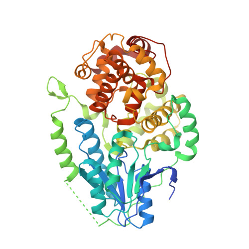
4CDM

Crystal structure of M. mazei photolyase soaked with synthetic 8-HDF


Experimental Data Snapshot

  • Method: X-RAY DIFFRACTION
  • Resolution: 2.70 Å
  • R-Value Free: 0.270 
  • R-Value Work: 0.206 
  • R-Value Observed: 0.207 

wwPDB Validation   3D Report Full Report


Ligand Structure Quality Assessment 


This is version 1.3 of the entry. See complete history


Literature

Structural and Evolutionary Aspects of Antenna Chromophore Usage by Class II Photolyases.

Kiontke, S.Gnau, P.Haselsberger, R.Batschauer, A.Essen, L.

(2014) J Biol Chem 289: 19659

  • DOI: https://doi.org/10.1074/jbc.M113.542431
  • Primary Citation of Related Structures:  
    4CDM, 4CDN

  • PubMed Abstract: 

    Light-harvesting and resonance energy transfer to the catalytic FAD cofactor are key roles for the antenna chromophores of light-driven DNA photolyases, which remove UV-induced DNA lesions. So far, five chemically diverse chromophores have been described for several photolyases and related cryptochromes, but no correlation between phylogeny and used antenna has been found. Despite a common protein topology, structural analysis of the distantly related class II photolyase from the archaeon Methanosarcina mazei (MmCPDII) as well as plantal orthologues indicated several differences in terms of DNA and FAD binding and electron transfer pathways. For MmCPDII we identify 8-hydroxydeazaflavin (8-HDF) as cognate antenna by in vitro and in vivo reconstitution, whereas the higher plant class II photolyase from Arabidopsis thaliana fails to bind any of the known chromophores. According to the 1.9 Å structure of the MmCPDII·8-HDF complex, its antenna binding site differs from other members of the photolyase-cryptochrome superfamily by an antenna loop that changes its conformation by 12 Å upon 8-HDF binding. Additionally, so-called N- and C-motifs contribute as conserved elements to the binding of deprotonated 8-HDF and allow predicting 8-HDF binding for most of the class II photolyases in the whole phylome. The 8-HDF antenna is used throughout the viridiplantae ranging from green microalgae to bryophyta and pteridophyta, i.e. mosses and ferns, but interestingly not in higher plants. Overall, we suggest that 8-hydroxydeazaflavin is a crucial factor for the survival of most higher eukaryotes which depend on class II photolyases to struggle with the genotoxic effects of solar UV exposure.


  • Organizational Affiliation

    From the Biomedical Research Centre/FB15, Unit for Structural Biochemistry, Philipps-University, Hans-Meerwein-Strasse, D-35032 Marburg, Germany.


Macromolecules
Find similar proteins by:  (by identity cutoff)  |  3D Structure
Entity ID: 1
MoleculeChains Sequence LengthOrganismDetailsImage
DEOXYRIBODIPYRIMIDINE PHOTOLYASE482Methanosarcina mazei Go1Mutation(s): 0 
EC: 4.1.99.3
UniProt
Find proteins for Q8PYK9 (Methanosarcina mazei (strain ATCC BAA-159 / DSM 3647 / Goe1 / Go1 / JCM 11833 / OCM 88))
Explore Q8PYK9 
Go to UniProtKB:  Q8PYK9
Entity Groups  
Sequence Clusters30% Identity50% Identity70% Identity90% Identity95% Identity100% Identity
UniProt GroupQ8PYK9
Sequence Annotations
Expand
  • Reference Sequence
Small Molecules
Ligands 4 Unique
IDChains Name / Formula / InChI Key2D Diagram3D Interactions
FAD
Query on FAD

Download Ideal Coordinates CCD File 
B [auth A]FLAVIN-ADENINE DINUCLEOTIDE
C27 H33 N9 O15 P2
VWWQXMAJTJZDQX-UYBVJOGSSA-N
FO1
Query on FO1

Download Ideal Coordinates CCD File 
C [auth A]1-deoxy-1-(8-hydroxy-2,4-dioxo-3,4-dihydropyrimido[4,5-b]quinolin-10(2H)-yl)-D-ribitol
C16 H17 N3 O7
AUEILLWDYUBWCM-XQQFMLRXSA-N
SO4
Query on SO4

Download Ideal Coordinates CCD File 
D [auth A],
E [auth A],
F [auth A],
G [auth A]
SULFATE ION
O4 S
QAOWNCQODCNURD-UHFFFAOYSA-L
GOL
Query on GOL

Download Ideal Coordinates CCD File 
H [auth A],
I [auth A]
GLYCEROL
C3 H8 O3
PEDCQBHIVMGVHV-UHFFFAOYSA-N
Experimental Data & Validation

Experimental Data

  • Method: X-RAY DIFFRACTION
  • Resolution: 2.70 Å
  • R-Value Free: 0.270 
  • R-Value Work: 0.206 
  • R-Value Observed: 0.207 
  • Space Group: P 43 21 2
Unit Cell:
Length ( Å )Angle ( ˚ )
a = 69.89α = 90
b = 69.89β = 90
c = 245.52γ = 90
Software Package:
Software NamePurpose
REFMACrefinement
XDSdata reduction
XSCALEdata scaling

Structure Validation

View Full Validation Report


Ligand Structure Quality Assessment 


Entry History 

Deposition Data

Revision History  (Full details and data files)

  • Version 1.0: 2014-05-21
    Type: Initial release
  • Version 1.1: 2014-06-04
    Changes: Database references
  • Version 1.2: 2014-08-13
    Changes: Database references
  • Version 1.3: 2024-05-08
    Changes: Data collection, Database references, Derived calculations, Other