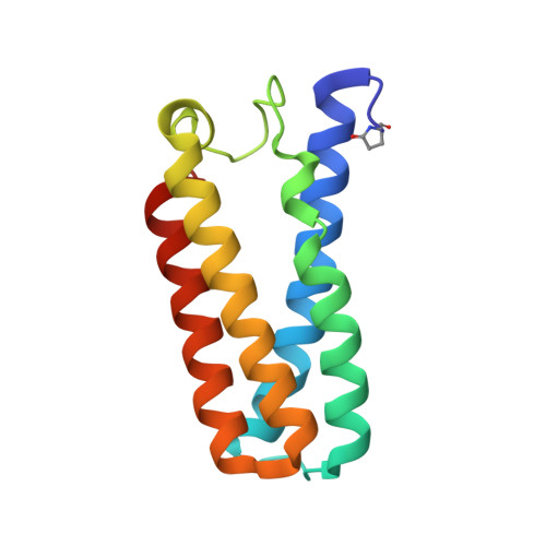
4CDY

Spectroscopically-validated structure of cytochrome c prime from Alcaligenes xylosoxidans, reduced by X-ray irradiation at 160K


Experimental Data Snapshot

  • Method: X-RAY DIFFRACTION
  • Resolution: 1.77 Å
  • R-Value Free: 0.239 
  • R-Value Work: 0.204 
  • R-Value Observed: 0.206 

Starting Model: experimental
View more details

wwPDB Validation   3D Report Full Report


Ligand Structure Quality Assessment 


This is version 2.1 of the entry. See complete history


Literature

Fingerprinting Redox and Ligand States in Haemprotein Crystal Structures Using Resonance Raman Spectroscopy.

Kekilli, D.Dworkowski, F.S.Pompidor, G.Fuchs, M.R.Andrew, C.R.Antonyuk, S.Strange, R.W.Eady, R.R.Hasnain, S.S.Hough, M.A.

(2014) Acta Crystallogr D Biol Crystallogr 70: 1289

  • DOI: https://doi.org/10.1107/S1399004714004039
  • Primary Citation of Related Structures:  
    4CDA, 4CDV, 4CDY, 4CIP, 4CJG, 4CJO

  • PubMed Abstract: 

    It is crucial to assign the correct redox and ligand states to crystal structures of proteins with an active redox centre to gain valid functional information and prevent the misinterpretation of structures. Single-crystal spectroscopies, particularly when applied in situ at macromolecular crystallography beamlines, allow spectroscopic investigations of redox and ligand states and the identification of reaction intermediates in protein crystals during the collection of structural data. Single-crystal resonance Raman spectroscopy was carried out in combination with macromolecular crystallography on Swiss Light Source beamline X10SA using cytochrome c' from Alcaligenes xylosoxidans. This allowed the fingerprinting and validation of different redox and ligand states, identification of vibrational modes and identification of intermediates together with monitoring of radiation-induced changes. This combined approach provides a powerful tool to obtain complementary data and correctly assign the true oxidation and ligand state(s) in redox-protein crystals.


  • Organizational Affiliation

    School of Biological Sciences, University of Essex, Wivenhoe Park, Colchester CO4 3SQ, England.


Macromolecules
Find similar proteins by:  (by identity cutoff)  |  3D Structure
Entity ID: 1
MoleculeChains Sequence LengthOrganismDetailsImage
CYTOCHROME C'127Achromobacter xylosoxidansMutation(s): 0 
UniProt
Find proteins for P00138 (Alcaligenes xylosoxydans xylosoxydans)
Explore P00138 
Go to UniProtKB:  P00138
Entity Groups  
Sequence Clusters30% Identity50% Identity70% Identity90% Identity95% Identity100% Identity
UniProt GroupP00138
Sequence Annotations
Expand
  • Reference Sequence
Small Molecules
Ligands 1 Unique
IDChains Name / Formula / InChI Key2D Diagram3D Interactions
HEC
Query on HEC

Download Ideal Coordinates CCD File 
B [auth A]HEME C
C34 H34 Fe N4 O4
HXQIYSLZKNYNMH-LJNAALQVSA-N
Modified Residues  1 Unique
IDChains TypeFormula2D DiagramParent
PCA
Query on PCA
A
L-PEPTIDE LINKINGC5 H7 N O3GLN
Experimental Data & Validation

Experimental Data

  • Method: X-RAY DIFFRACTION
  • Resolution: 1.77 Å
  • R-Value Free: 0.239 
  • R-Value Work: 0.204 
  • R-Value Observed: 0.206 
  • Space Group: P 65 2 2
Unit Cell:
Length ( Å )Angle ( ˚ )
a = 53.623α = 90
b = 53.623β = 90
c = 181.724γ = 120
Software Package:
Software NamePurpose
REFMACrefinement
XDSdata reduction
SCALAdata scaling

Structure Validation

View Full Validation Report


Ligand Structure Quality Assessment 


Entry History 

Deposition Data

Revision History  (Full details and data files)

  • Version 1.0: 2014-05-21
    Type: Initial release
  • Version 2.0: 2020-03-11
    Changes: Advisory, Derived calculations, Other, Polymer sequence
  • Version 2.1: 2023-12-20
    Changes: Advisory, Data collection, Database references, Derived calculations, Refinement description