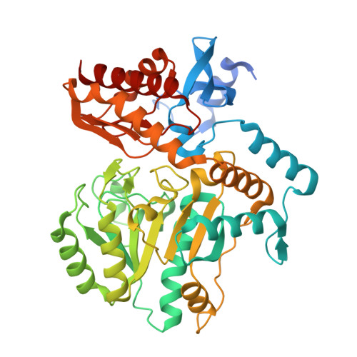
4CXQ

Mycobaterium tuberculosis transaminase BioA complexed with substrate KAPA


Experimental Data Snapshot

  • Method: X-RAY DIFFRACTION
  • Resolution: 1.80 Å
  • R-Value Free: 0.195 
  • R-Value Work: 0.155 
  • R-Value Observed: 0.157 

Starting Model: experimental
View more details

wwPDB Validation   3D Report Full Report


Ligand Structure Quality Assessment 


This is version 1.3 of the entry. See complete history


Literature

Inhibition of Mycobacterium Tuberculosis Transaminase Bioa by Aryl Hydrazines and Hydrazides.

Dai, R.Wilson, D.J.Geders, T.W.Aldrich, C.C.Finzel, B.C.

(2014) Chembiochem 15: 575

  • DOI: https://doi.org/10.1002/cbic.201300748
  • Primary Citation of Related Structures:  
    4CXQ, 4CXR, 4MQP, 4MQQ, 4MQR

  • PubMed Abstract: 

    7,8-Diaminopelargonic acid synthase (BioA) of Mycobacterium tuberculosis is a recently validated target for therapeutic intervention in the treatment of tuberculosis (TB). Using biophysical fragment screening and structural characterization of compounds, we have identified a potent aryl hydrazine inhibitor of BioA that reversibly modifies the pyridoxal-5'-phosphate (PLP) cofactor, forming a stable quinonoid. Analogous hydrazides also form covalent adducts that can be observed crystallographically but are incapable of inactivating the enzyme. In the X-ray crystal structures, small molecules induce unexpected conformational remodeling in the substrate binding site. We compared these conformational changes to those induced upon binding of the substrate (7-keto-8-aminopelargonic acid), and characterized the inhibition kinetics and the X-ray crystal structures of BioA with the hydrazine compound and analogues to unveil the mechanism of this reversible covalent modification.


  • Organizational Affiliation

    Department of Medicinal Chemistry, University of Minnesota, 308 Harvard St. SE, Minneapolis, MN 55455 (USA).


Macromolecules
Find similar proteins by:  (by identity cutoff)  |  3D Structure
Entity ID: 1
MoleculeChains Sequence LengthOrganismDetailsImage
ADENOSYLMETHIONINE-8-AMINO-7-OXONONANOATE AMINOTRANSFERASE
A, B
457Mycobacterium tuberculosisMutation(s): 0 
EC: 2.6.1.62
UniProt
Find proteins for P9WQ81 (Mycobacterium tuberculosis (strain ATCC 25618 / H37Rv))
Explore P9WQ81 
Go to UniProtKB:  P9WQ81
Entity Groups  
Sequence Clusters30% Identity50% Identity70% Identity90% Identity95% Identity100% Identity
UniProt GroupP9WQ81
Sequence Annotations
Expand
  • Reference Sequence
Small Molecules
Ligands 4 Unique
IDChains Name / Formula / InChI Key2D Diagram3D Interactions
PLP
Query on PLP

Download Ideal Coordinates CCD File 
C [auth A],
H [auth B]
PYRIDOXAL-5'-PHOSPHATE
C8 H10 N O6 P
NGVDGCNFYWLIFO-UHFFFAOYSA-N
KAP
Query on KAP

Download Ideal Coordinates CCD File 
D [auth A],
I [auth B]
7-KETO-8-AMINOPELARGONIC ACID
C9 H17 N O3
GUAHPAJOXVYFON-ZETCQYMHSA-N
PEG
Query on PEG

Download Ideal Coordinates CCD File 
E [auth A],
J [auth B]
DI(HYDROXYETHYL)ETHER
C4 H10 O3
MTHSVFCYNBDYFN-UHFFFAOYSA-N
EDO
Query on EDO

Download Ideal Coordinates CCD File 
F [auth A],
G [auth A],
K [auth B]
1,2-ETHANEDIOL
C2 H6 O2
LYCAIKOWRPUZTN-UHFFFAOYSA-N
Experimental Data & Validation

Experimental Data

  • Method: X-RAY DIFFRACTION
  • Resolution: 1.80 Å
  • R-Value Free: 0.195 
  • R-Value Work: 0.155 
  • R-Value Observed: 0.157 
  • Space Group: P 21 21 21
Unit Cell:
Length ( Å )Angle ( ˚ )
a = 62.553α = 90
b = 66.149β = 90
c = 205.294γ = 90
Software Package:
Software NamePurpose
PHENIXrefinement
XDSdata reduction
SCALAdata scaling
PHASERphasing

Structure Validation

View Full Validation Report


Ligand Structure Quality Assessment 


Entry History 

Deposition Data

Revision History  (Full details and data files)

  • Version 1.0: 2014-04-23
    Type: Initial release
  • Version 1.1: 2019-01-30
    Changes: Data collection, Experimental preparation, Other
  • Version 1.2: 2019-02-06
    Changes: Data collection, Experimental preparation
  • Version 1.3: 2023-12-20
    Changes: Data collection, Database references, Derived calculations, Other, Refinement description