4E2Q | pdb_00004e2q

Crystal Structure of (S)-Ureidoglycine Aminohydrolase from Arabidopsis thaliana


Experimental Data Snapshot

  • Method: X-RAY DIFFRACTION
  • Resolution: 2.50 Å
  • R-Value Free: 
    0.286 (Depositor), 0.244 (DCC) 
  • R-Value Work: 
    0.224 (Depositor), 0.242 (DCC) 

wwPDB Validation 3D Report Full Report

Validation slider image for 4E2Q

This is version 1.2 of the entry. See complete history

Literature

Structural and functional insights into (S)-ureidoglycine aminohydrolase, key enzyme of purine catabolism in Arabidopsis thaliana

Shin, I.Percudani, R.Rhee, S.

(2012) J Biological Chem 287: 18796-18805

  • DOI: https://doi.org/10.1074/jbc.M111.331819
  • Primary Citation Related Structures: 
    4E2Q, 4E2S

  • PubMed Abstract: 

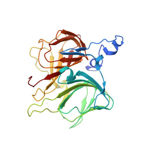
    The ureide pathway has recently been identified as the metabolic route of purine catabolism in plants and some bacteria. In this pathway, uric acid, which is a major product of the early stage of purine catabolism, is degraded into glyoxylate and ammonia via stepwise reactions of seven different enzymes. Therefore, the pathway has a possible physiological role in mobilization of purine ring nitrogen for further assimilation. (S)-Ureidoglycine aminohydrolase enzyme converts (S)-ureidoglycine into (S)-ureidoglycolate and ammonia, providing the final substrate to the pathway. Here, we report a structural and functional analysis of this enzyme from Arabidopsis thaliana (AtUGlyAH). The crystal structure of AtUGlyAH in the ligand-free form shows a monomer structure in the bicupin fold of the β-barrel and an octameric functional unit as well as a Mn(2+) ion binding site. The structure of AtUGlyAH in complex with (S)-ureidoglycine revealed that the Mn(2+) ion acts as a molecular anchor to bind (S)-ureidoglycine, and its binding mode dictates the enantioselectivity of the reaction. Further kinetic analysis characterized the functional roles of the active site residues, including the Mn(2+) ion binding site and residues in the vicinity of (S)-ureidoglycine. These analyses provide molecular insights into the structure of the enzyme and its possible catalytic mechanism.


  • Organizational Affiliation
    • Department of Agricultural Biotechnology, Seoul National University, Seoul, Korea.

Macromolecule Content 

  • Total Structure Weight: 484.55 kDa 
  • Atom Count: 33,964 
  • Modeled Residue Count: 4,128 
  • Deposited Residue Count: 4,256 
  • Unique protein chains: 1

Macromolecules

Find similar proteins by:|  3D Structure
Entity ID: 1
MoleculeChains  Sequence LengthOrganismDetailsImage
Ureidoglycine aminohydrolase
A, B, C, D, E
A, B, C, D, E, F, G, H, I, J, K, L, M, N, O, P
266Arabidopsis thalianaMutation(s): 0 
Gene Names: ylbAUGLYAHAt4g17050
EC: 3.5.3 (PDB Primary Data), 3.5.3.26 (UniProt)
UniProt
Find proteins for Q8GXV5 (Arabidopsis thaliana)
Explore Q8GXV5 
Go to UniProtKB:  Q8GXV5
Entity Groups
Sequence Clusters30% Identity50% Identity70% Identity90% Identity95% Identity100% Identity
UniProt GroupQ8GXV5
Sequence Annotations
Expand
Reference Sequence

Small Molecules

Ligands 1 Unique
IDChains Name / Formula / InChI Key2D Diagram3D Interactions
MN

Query on MN


Download:Ideal Coordinates CCD File
AA [auth K]
BA [auth L]
CA [auth M]
DA [auth N]
EA [auth O]
AA [auth K],
BA [auth L],
CA [auth M],
DA [auth N],
EA [auth O],
FA [auth P],
Q [auth A],
R [auth B],
S [auth C],
T [auth D],
U [auth E],
V [auth F],
W [auth G],
X [auth H],
Y [auth I],
Z [auth J]
MANGANESE (II) ION
Mn
WAEMQWOKJMHJLA-UHFFFAOYSA-N

Experimental Data & Validation

Experimental Data

  • Method: X-RAY DIFFRACTION
  • Resolution: 2.50 Å
  • R-Value Free:  0.286 (Depositor), 0.244 (DCC) 
  • R-Value Work:  0.224 (Depositor), 0.242 (DCC) 
Space Group: P 1 21 1
Unit Cell:
Length ( Å )Angle ( ˚ )
a = 93.856α = 90
b = 175.773β = 99.39
c = 155.176γ = 90
Software Package:
Software NamePurpose
HKL-2000data collection
PHENIXmodel building
CNSrefinement
HKL-2000data reduction
HKL-2000data scaling
PHENIXphasing

Structure Validation

View Full Validation Report


Entry History 

Deposition Data

Revision History  (Full details and data files)

  • Version 1.0: 2012-04-18
    Type: Initial release
  • Version 1.1: 2013-07-10
    Changes: Database references
  • Version 1.2: 2024-03-20
    Changes: Data collection, Database references, Derived calculations