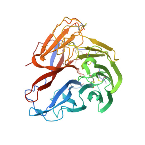
4GDI

A subtype N10 neuraminidase-like protein of A/little yellow-shouldered bat/Guatemala/164/2009


Experimental Data Snapshot

  • Method: X-RAY DIFFRACTION
  • Resolution: 1.95 Å
  • R-Value Free: 0.223 
  • R-Value Work: 0.180 
  • R-Value Observed: 0.182 

Starting Model: experimental
View more details

wwPDB Validation   3D Report Full Report


Ligand Structure Quality Assessment 


This is version 2.2 of the entry. See complete history


Literature

Crystal structures of two subtype N10 neuraminidase-like proteins from bat influenza A viruses reveal a diverged putative active site.

Zhu, X.Yang, H.Guo, Z.Yu, W.Carney, P.J.Li, Y.Chen, L.M.Paulson, J.C.Donis, R.O.Tong, S.Stevens, J.Wilson, I.A.

(2012) Proc Natl Acad Sci U S A 109: 18903-18908

  • DOI: https://doi.org/10.1073/pnas.1212579109
  • Primary Citation of Related Structures:  
    4GDI, 4GDJ, 4GEZ

  • PubMed Abstract: 

    Recently, we reported a unique influenza A virus subtype H17N10 from little yellow-shouldered bats. Its neuraminidase (NA) gene encodes a protein that appears to be highly divergent from all known influenza NAs and was assigned as a new subtype N10. To provide structural and functional insights on the bat H17N10 virus, X-ray structures were determined for N10 NA proteins from influenza A viruses A/little yellow-shouldered bat/Guatemala/164/2009 (GU09-164) in two crystal forms at 1.95 Å and 2.5 Å resolution and A/little yellow-shouldered bat/Guatemala/060/2010 (GU10-060) at 2.0 Å. The overall N10 structures are similar to each other and to other known influenza NA structures, with a single highly conserved calcium binding site in each monomer. However, the region corresponding to the highly conserved active site of influenza A N1-N9 NA subtypes and influenza B NA differs substantially. In particular, most of the amino acid residues required for NA activity are substituted, and the putative active site is much wider because of displacement of the 150-loop and 430-loop. These structural features and the fact that the recombinant N10 protein exhibits no, or extremely low, NA activity suggest that it may have a different function than the NA proteins of other influenza viruses. Accordingly, we propose that the N10 protein be termed an NA-like protein until its function is elucidated.


  • Organizational Affiliation

    Department of Molecular Biology, The Scripps Research Institute, La Jolla, CA 92037, USA.


Macromolecules
Find similar proteins by:  (by identity cutoff)  |  3D Structure
Entity ID: 1
MoleculeChains Sequence LengthOrganismDetailsImage
Neuraminidase
A, B, C, D, E
A, B, C, D, E, F
373Influenza A virus (A/little yellow-shouldered bat/Guatemala/164/2009(H17N10))Mutation(s): 0 
Gene Names: NA
EC: 3.2.1.18
UniProt
Find proteins for H6QM85 (Influenza A virus)
Explore H6QM85 
Go to UniProtKB:  H6QM85
Entity Groups  
Sequence Clusters30% Identity50% Identity70% Identity90% Identity95% Identity100% Identity
UniProt GroupH6QM85
Glycosylation
Glycosylation Sites: 4
Sequence Annotations
Expand
  • Reference Sequence
Oligosaccharides

Help

Entity ID: 2
MoleculeChains Length2D Diagram Glycosylation3D Interactions
beta-D-mannopyranose-(1-4)-2-acetamido-2-deoxy-beta-D-glucopyranose-(1-4)-[beta-L-fucopyranose-(1-3)][alpha-L-fucopyranose-(1-6)]2-acetamido-2-deoxy-beta-D-glucopyranose
G
5N-Glycosylation
Glycosylation Resources
GlyTouCan:  G31349EA
GlyCosmos:  G31349EA
Entity ID: 3
MoleculeChains Length2D Diagram Glycosylation3D Interactions
alpha-D-mannopyranose-(1-3)-beta-D-mannopyranose-(1-4)-2-acetamido-2-deoxy-beta-D-glucopyranose-(1-4)-[beta-L-fucopyranose-(1-3)][alpha-L-fucopyranose-(1-6)]2-acetamido-2-deoxy-beta-D-glucopyranose
H
6N-Glycosylation
Glycosylation Resources
GlyTouCan:  G88708XF
GlyCosmos:  G88708XF
GlyGen:  G88708XF
Entity ID: 4
MoleculeChains Length2D Diagram Glycosylation3D Interactions
beta-L-fucopyranose-(1-3)-[2-acetamido-2-deoxy-beta-D-glucopyranose-(1-4)][alpha-L-fucopyranose-(1-6)]2-acetamido-2-deoxy-beta-D-glucopyranose
I, K, M
4N-Glycosylation
Glycosylation Resources
GlyTouCan:  G62810GW
GlyCosmos:  G62810GW
Entity ID: 5
MoleculeChains Length2D Diagram Glycosylation3D Interactions
2-acetamido-2-deoxy-beta-D-glucopyranose-(1-4)-[alpha-L-fucopyranose-(1-6)]2-acetamido-2-deoxy-beta-D-glucopyranose
J
3N-Glycosylation
Glycosylation Resources
GlyTouCan:  G21290RB
GlyCosmos:  G21290RB
GlyGen:  G21290RB
Entity ID: 6
MoleculeChains Length2D Diagram Glycosylation3D Interactions
alpha-L-fucopyranose-(1-6)-2-acetamido-2-deoxy-beta-D-glucopyranose
L
2N-Glycosylation
Glycosylation Resources
GlyTouCan:  G86851RC
GlyCosmos:  G86851RC
GlyGen:  G86851RC
Entity ID: 7
MoleculeChains Length2D Diagram Glycosylation3D Interactions
beta-L-fucopyranose-(1-3)-[2-acetamido-2-deoxy-beta-D-glucopyranose-(1-4)]2-acetamido-2-deoxy-beta-D-glucopyranose
N
3N-Glycosylation
Glycosylation Resources
GlyTouCan:  G46874CY
GlyCosmos:  G46874CY
GlyGen:  G46874CY
Small Molecules
Ligands 4 Unique
IDChains Name / Formula / InChI Key2D Diagram3D Interactions
NAG
Query on NAG

Download Ideal Coordinates CCD File 
DB [auth F]
EB [auth F]
IA [auth C]
JA [auth C]
O [auth A]
DB [auth F],
EB [auth F],
IA [auth C],
JA [auth C],
O [auth A],
P [auth A],
PA [auth D],
Q [auth A],
QA [auth D],
WA [auth E],
X [auth B],
XA [auth E],
Y [auth B]
2-acetamido-2-deoxy-beta-D-glucopyranose
C8 H15 N O6
OVRNDRQMDRJTHS-FMDGEEDCSA-N
GOL
Query on GOL

Download Ideal Coordinates CCD File 
AB [auth E]
BA [auth B]
BB [auth E]
CA [auth B]
CB [auth E]
AB [auth E],
BA [auth B],
BB [auth E],
CA [auth B],
CB [auth E],
HB [auth F],
IB [auth F],
JB [auth F],
KB [auth F],
MA [auth C],
S [auth A],
T [auth A],
TA [auth D],
U [auth A]
GLYCEROL
C3 H8 O3
PEDCQBHIVMGVHV-UHFFFAOYSA-N
NO3
Query on NO3

Download Ideal Coordinates CCD File 
DA [auth B]
EA [auth B]
FA [auth B]
GA [auth B]
HA [auth B]
DA [auth B],
EA [auth B],
FA [auth B],
GA [auth B],
HA [auth B],
LB [auth F],
MB [auth F],
NA [auth C],
NB [auth F],
OA [auth C],
UA [auth D],
V [auth A],
VA [auth D],
W [auth A]
NITRATE ION
N O3
NHNBFGGVMKEFGY-UHFFFAOYSA-N
CA
Query on CA

Download Ideal Coordinates CCD File 
AA [auth B]
FB [auth F]
GB [auth F]
KA [auth C]
LA [auth C]
AA [auth B],
FB [auth F],
GB [auth F],
KA [auth C],
LA [auth C],
R [auth A],
RA [auth D],
SA [auth D],
YA [auth E],
Z [auth B],
ZA [auth E]
CALCIUM ION
Ca
BHPQYMZQTOCNFJ-UHFFFAOYSA-N
Experimental Data & Validation

Experimental Data

  • Method: X-RAY DIFFRACTION
  • Resolution: 1.95 Å
  • R-Value Free: 0.223 
  • R-Value Work: 0.180 
  • R-Value Observed: 0.182 
  • Space Group: I 4
Unit Cell:
Length ( Å )Angle ( ˚ )
a = 176.324α = 90
b = 176.324β = 90
c = 193.313γ = 90
Software Package:
Software NamePurpose
PHENIXrefinement

Structure Validation

View Full Validation Report


Ligand Structure Quality Assessment 


Entry History 

Deposition Data

Revision History  (Full details and data files)

  • Version 1.0: 2012-09-26
    Type: Initial release
  • Version 1.1: 2012-10-10
    Changes: Database references
  • Version 1.2: 2012-11-28
    Changes: Database references
  • Version 1.3: 2014-12-17
    Changes: Data collection
  • Version 2.0: 2020-07-29
    Type: Remediation
    Reason: Carbohydrate remediation
    Changes: Atomic model, Data collection, Database references, Derived calculations, Structure summary
  • Version 2.1: 2023-09-13
    Changes: Data collection, Database references, Refinement description, Structure summary
  • Version 2.2: 2024-10-16
    Changes: Structure summary