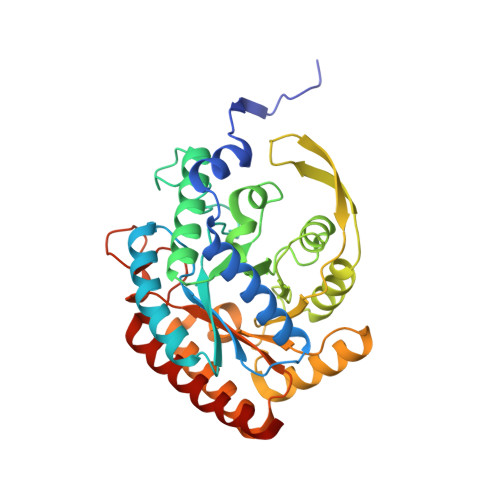
4HSO

Crystal structure of S213G variant DAH7PS from Neisseria meningitidis


Experimental Data Snapshot

  • Method: X-RAY DIFFRACTION
  • Resolution: 2.10 Å
  • R-Value Free: 0.242 
  • R-Value Work: 0.204 
  • R-Value Observed: 0.206 

Starting Model: experimental
View more details

wwPDB Validation   3D Report Full Report


Ligand Structure Quality Assessment 


This is version 1.1 of the entry. See complete history


Literature

Neisseria meningitidis expresses a single 3-deoxy-d-arabino-heptulosonate 7-phosphate synthase that is inhibited primarily by phenylalanine.

Cross, P.J.Pietersma, A.L.Allison, T.M.Wilson-Coutts, S.M.Cochrane, F.C.Parker, E.J.

(2013) Protein Sci 22: 1087-1099

  • DOI: https://doi.org/10.1002/pro.2293
  • Primary Citation of Related Structures:  
    4HSN, 4HSO, 4IXX

  • PubMed Abstract: 

    Neisseria meningitidis is the causative agent of meningitis and meningococcal septicemia is a major cause of disease worldwide, resulting in brain damage and hearing loss, and can be fatal in a large proportion of cases. The enzyme 3-deoxy-d-arabino-heptulosonate 7-phosphate synthase (DAH7PS) catalyzes the first reaction in the shikimate pathway leading to the biosynthesis of aromatic metabolites including the aromatic acids l-Trp, l-Phe, and l-Tyr. This pathway is absent in humans, meaning that enzymes of the pathway are considered as potential candidates for therapeutic intervention. As the entry point, feedback inhibition of DAH7PS by pathway end products is a key mechanism for the control of pathway flux. The structure of the single DAH7PS expressed by N. meningitidis was determined at 2.0 Å resolution. In contrast to the other DAH7PS enzymes, which are inhibited only by a single aromatic amino acid, the N. meningitidis DAH7PS was inhibited by all three aromatic amino acids, showing greatest sensitivity to l-Phe. An N. meningitidis enzyme variant, in which a single Ser residue at the bottom of the inhibitor-binding cavity was substituted to Gly, altered inhibitor specificity from l-Phe to l-Tyr. Comparison of the crystal structures of both unbound and Tyr-bound forms and the small angle X-ray scattering profiles reveal that N. meningtidis DAH7PS undergoes no significant conformational change on inhibitor binding. These observations are consistent with an allosteric response arising from changes in protein motion rather than conformation, and suggest ligands that modulate protein dynamics may be effective inhibitors of this enzyme.


  • Organizational Affiliation

    Biomolecular Interaction Centre and Department of Chemistry, University of Canterbury, Christchurch, New Zealand.


Macromolecules
Find similar proteins by:  (by identity cutoff)  |  3D Structure
Entity ID: 1
MoleculeChains Sequence LengthOrganismDetailsImage
3-deoxy-D-arabino-heptulosonate 7-phosphate synthase
A, B, C, D
351Neisseria meningitidis MC58Mutation(s): 1 
Gene Names: aroGNMB0307
EC: 2.5.1.54
UniProt
Find proteins for Q9K169 (Neisseria meningitidis serogroup B (strain ATCC BAA-335 / MC58))
Explore Q9K169 
Go to UniProtKB:  Q9K169
Entity Groups  
Sequence Clusters30% Identity50% Identity70% Identity90% Identity95% Identity100% Identity
UniProt GroupQ9K169
Sequence Annotations
Expand
  • Reference Sequence
Small Molecules
Ligands 3 Unique
IDChains Name / Formula / InChI Key2D Diagram3D Interactions
TYR
Query on TYR

Download Ideal Coordinates CCD File 
E [auth A],
H [auth B],
K [auth C],
N [auth D]
TYROSINE
C9 H11 N O3
OUYCCCASQSFEME-QMMMGPOBSA-N
PEP
Query on PEP

Download Ideal Coordinates CCD File 
F [auth A],
I [auth B],
L [auth C],
O [auth D]
PHOSPHOENOLPYRUVATE
C3 H5 O6 P
DTBNBXWJWCWCIK-UHFFFAOYSA-N
MN
Query on MN

Download Ideal Coordinates CCD File 
G [auth A],
J [auth B],
M [auth C],
P [auth D]
MANGANESE (II) ION
Mn
WAEMQWOKJMHJLA-UHFFFAOYSA-N
Binding Affinity Annotations 
IDSourceBinding Affinity
TYR PDBBind:  4HSO Kd: 2.20e+4 (nM) from 1 assay(s)
Experimental Data & Validation

Experimental Data

  • Method: X-RAY DIFFRACTION
  • Resolution: 2.10 Å
  • R-Value Free: 0.242 
  • R-Value Work: 0.204 
  • R-Value Observed: 0.206 
  • Space Group: P 1 21 1
Unit Cell:
Length ( Å )Angle ( ˚ )
a = 73.523α = 90
b = 136.995β = 96.63
c = 76.212γ = 90
Software Package:
Software NamePurpose
SCALAdata scaling
REFMACrefinement
PDB_EXTRACTdata extraction
XSCALEdata scaling

Structure Validation

View Full Validation Report


Ligand Structure Quality Assessment 


Entry History 

Deposition Data

Revision History  (Full details and data files)

  • Version 1.0: 2013-09-11
    Type: Initial release
  • Version 1.1: 2023-09-20
    Changes: Data collection, Database references, Derived calculations, Refinement description