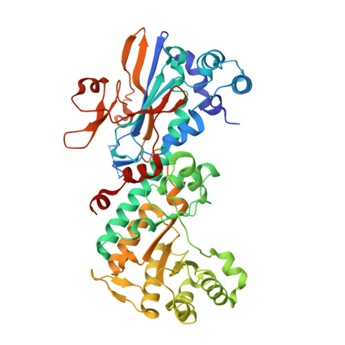
4L4L

Structural Analysis of a Phosphoribosylated Inhibitor in Complex with Human Nicotinamide Phosphoribosyltransferase


Experimental Data Snapshot

  • Method: X-RAY DIFFRACTION
  • Resolution: 2.12 Å
  • R-Value Free: 0.227 
  • R-Value Work: 0.187 
  • R-Value Observed: 0.189 

wwPDB Validation   3D Report Full Report


Ligand Structure Quality Assessment 


This is version 1.2 of the entry. See complete history


Literature

Structural and biochemical analyses of the catalysis and potency impact of inhibitor phosphoribosylation by human nicotinamide phosphoribosyltransferase.

Oh, A.Ho, Y.C.Zak, M.Liu, Y.Chen, X.Yuen, P.W.Zheng, X.Liu, Y.Dragovich, P.S.Wang, W.

(2014) Chembiochem 15: 1121-1130

  • DOI: https://doi.org/10.1002/cbic.201402023
  • Primary Citation of Related Structures:  
    4L4L, 4L4M, 4O0Z, 4O10, 4O12

  • PubMed Abstract: 

    Prolonged inhibition of nicotinamide phosphoribosyltransferase (NAMPT) is a strategy for targeting cancer metabolism. Many NAMPT inhibitors undergo NAMPT-catalyzed phosphoribosylation (pRib), a property often correlated with their cellular potency. To understand this phenomenon and facilitate drug design, we analyzed a potent cellularly active NAMPT inhibitor (GNE-617). A crystal structure of pRib-GNE-617 in complex with NAMPT protein revealed a relaxed binding mode. Consistently, the adduct formation resulted in tight binding and strong product inhibition. In contrast, a biochemically equipotent isomer of GNE-617 (GNE-643) also formed pRib adducts but displayed significantly weaker cytotoxicity. Structural analysis revealed an altered ligand conformation of GNE-643, thus suggesting weak association of the adducts with NAMPT. Our data support a model for cellularly active NAMPT inhibitors that undergo NAMPT-catalyzed phosphoribosylation to produce pRib adducts that retain efficient binding to the enzyme.


  • Organizational Affiliation

    Genentech, Inc., Department of Structural Biology, 1 DNA Way, South San Francisco, California 94080 (USA).


Macromolecules
Find similar proteins by:  (by identity cutoff)  |  3D Structure
Entity ID: 1
MoleculeChains Sequence LengthOrganismDetailsImage
Nicotinamide phosphoribosyltransferase
A, B
501Homo sapiensMutation(s): 0 
Gene Names: NAMPTPBEFPBEF1
EC: 2.4.2.12
UniProt & NIH Common Fund Data Resources
Find proteins for P43490 (Homo sapiens)
Explore P43490 
Go to UniProtKB:  P43490
PHAROS:  P43490
GTEx:  ENSG00000105835 
Entity Groups  
Sequence Clusters30% Identity50% Identity70% Identity90% Identity95% Identity100% Identity
UniProt GroupP43490
Sequence Annotations
Expand
  • Reference Sequence
Small Molecules
Ligands 3 Unique
IDChains Name / Formula / InChI Key2D Diagram3D Interactions
1XC
Query on 1XC

Download Ideal Coordinates CCD File 
C [auth A],
I [auth B]
6-({4-[(3,5-difluorophenyl)sulfonyl]benzyl}carbamoyl)-1-(5-O-phosphono-beta-D-ribofuranosyl)imidazo[1,2-a]pyridin-1-ium
C26 H25 F2 N3 O10 P S
ZYTGSQWLYZVDKX-IGGXFAESSA-O
PO4
Query on PO4

Download Ideal Coordinates CCD File 
D [auth A],
E [auth A],
J [auth B],
K [auth B]
PHOSPHATE ION
O4 P
NBIIXXVUZAFLBC-UHFFFAOYSA-K
EDO
Query on EDO

Download Ideal Coordinates CCD File 
F [auth A],
G [auth A],
H [auth A]
1,2-ETHANEDIOL
C2 H6 O2
LYCAIKOWRPUZTN-UHFFFAOYSA-N
Experimental Data & Validation

Experimental Data

  • Method: X-RAY DIFFRACTION
  • Resolution: 2.12 Å
  • R-Value Free: 0.227 
  • R-Value Work: 0.187 
  • R-Value Observed: 0.189 
  • Space Group: P 1 21 1
Unit Cell:
Length ( Å )Angle ( ˚ )
a = 60.215α = 90
b = 106.857β = 96.53
c = 83.021γ = 90
Software Package:
Software NamePurpose
HKL-2000data collection
PHASERphasing
PHENIXrefinement
DENZOdata reduction
SCALEPACKdata scaling

Structure Validation

View Full Validation Report


Ligand Structure Quality Assessment 


Entry History 

Deposition Data

Revision History  (Full details and data files)

  • Version 1.0: 2014-06-11
    Type: Initial release
  • Version 1.1: 2014-10-22
    Changes: Database references
  • Version 1.2: 2024-02-28
    Changes: Data collection, Database references, Derived calculations