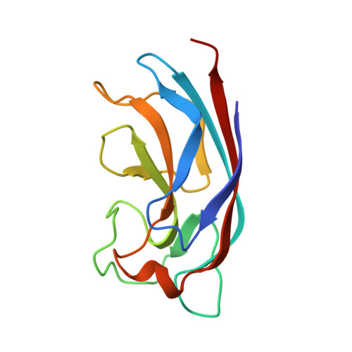
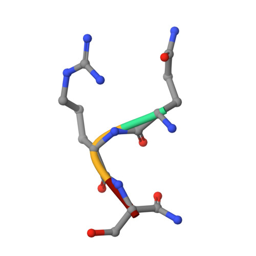
4LKD

Crystal Structure of Pseudomonas aeruginosa Lectin LecA Complexed with GalA-QRS at 2.31 A Resolution


Experimental Data Snapshot

  • Method: X-RAY DIFFRACTION
  • Resolution: 2.31 Å
  • R-Value Free: 0.247 
  • R-Value Work: 0.215 
  • R-Value Observed: 0.216 

wwPDB Validation   3D Report Full Report


Ligand Structure Quality Assessment 


This is version 1.2 of the entry. See complete history


Literature

Structure-Based Optimization of the Terminal Tripeptide in Glycopeptide Dendrimer Inhibitors of Pseudomonas aeruginosa Biofilms Targeting LecA.

Kadam, R.U.Bergmann, M.Garg, D.Gabrieli, G.Stocker, A.Darbre, T.Reymond, J.-L.

(2013) Chemistry 19: 17054-17063

  • DOI: https://doi.org/10.1002/chem.201302587
  • Primary Citation of Related Structures:  
    4LKD, 4LKE, 4LKF

  • PubMed Abstract: 

    The galactopeptide dendrimer GalAG2 ((β-Gal-OC6H4CO-Lys-Pro-Leu)4(Lys-Phe-Lys-Ile)2Lys-His-Ile-NH2) binds strongly to the Pseudomonas aeruginosa (PA) lectin LecA, and it inhibits PA biofilms, as well as disperses already established ones. By starting with the crystal structure of the terminal tripeptide moiety GalA-KPL in complex with LecA, a computational mutagenesis study was carried out on the galactotripeptide to optimize the peptide-lectin interactions. 25 mutants were experimentally evaluated by a hemagglutination inhibition assay, 17 by isothermal titration calorimetry, and 3 by X-ray crystallography. Two of these tripeptides, GalA-KPY (dissociation constant (K(D))=2.7 μM) and GalA-KRL (K(D)=2.7 μM), are among the most potent monovalent LecA ligands reported to date. Dendrimers based on these tripeptide ligands showed improved PA biofilm inhibition and dispersal compared to those of GalAG2, particularly G2KPY ((β-Gal-OC6H4CO-Lys-Pro-Tyr)4(Lys-Phe-Lys-Ile)2Lys-His-Ile-NH2). The possibility to retain and even improve the biofilm inhibition in several analogues of GalAG2 suggests that it should be possible to fine-tune this dendrimer towards therapeutic use by adjusting the pharmacokinetic parameters in addition to the biofilm inhibition through amino acid substitutions.


  • Organizational Affiliation

    Department of Chemistry and Biochemistry, University of Berne, Freiestrasse 3, 3012 Berne (Switzerland), Fax: (+41) 31-631-80-57.


Macromolecules
Find similar proteins by:  (by identity cutoff)  |  3D Structure
Entity ID: 1
MoleculeChains Sequence LengthOrganismDetailsImage
PA-I galactophilic lectin
A, B, C, D, E
A, B, C, D, E, F, G, H
121Pseudomonas aeruginosa PAO1Mutation(s): 0 
Gene Names: lecApa1LPA2570
UniProt
Find proteins for Q05097 (Pseudomonas aeruginosa (strain ATCC 15692 / DSM 22644 / CIP 104116 / JCM 14847 / LMG 12228 / 1C / PRS 101 / PAO1))
Explore Q05097 
Go to UniProtKB:  Q05097
Entity Groups  
Sequence Clusters30% Identity50% Identity70% Identity90% Identity95% Identity100% Identity
UniProt GroupQ05097
Sequence Annotations
Expand
  • Reference Sequence

Find similar proteins by:  Sequence   |   3D Structure  

Entity ID: 2
MoleculeChains Sequence LengthOrganismDetailsImage
peptide QRSA
I, J, K, L, M
I, J, K, L, M, N, O, P
4N/AMutation(s): 0 
Sequence Annotations
Expand
  • Reference Sequence
Small Molecules
Ligands 3 Unique
IDChains Name / Formula / InChI Key2D Diagram3D Interactions
GAL
Query on GAL

Download Ideal Coordinates CCD File 
AA [auth F]
CA [auth G]
EA [auth H]
Q [auth A]
S [auth B]
AA [auth F],
CA [auth G],
EA [auth H],
Q [auth A],
S [auth B],
U [auth C],
W [auth D],
Y [auth E]
beta-D-galactopyranose
C6 H12 O6
WQZGKKKJIJFFOK-FPRJBGLDSA-N
PHB
Query on PHB

Download Ideal Coordinates CCD File 
GA [auth I]
HA [auth J]
IA [auth K]
JA [auth L]
KA [auth M]
GA [auth I],
HA [auth J],
IA [auth K],
JA [auth L],
KA [auth M],
LA [auth N],
MA [auth O],
NA [auth P]
P-HYDROXYBENZOIC ACID
C7 H6 O3
FJKROLUGYXJWQN-UHFFFAOYSA-N
CA
Query on CA

Download Ideal Coordinates CCD File 
BA [auth F]
DA [auth G]
FA [auth H]
R [auth A]
T [auth B]
BA [auth F],
DA [auth G],
FA [auth H],
R [auth A],
T [auth B],
V [auth C],
X [auth D],
Z [auth E]
CALCIUM ION
Ca
BHPQYMZQTOCNFJ-UHFFFAOYSA-N
Experimental Data & Validation

Experimental Data

  • Method: X-RAY DIFFRACTION
  • Resolution: 2.31 Å
  • R-Value Free: 0.247 
  • R-Value Work: 0.215 
  • R-Value Observed: 0.216 
  • Space Group: C 1 2 1
Unit Cell:
Length ( Å )Angle ( ˚ )
a = 159.216α = 90
b = 149.239β = 110.96
c = 87.018γ = 90
Software Package:
Software NamePurpose
SLSPSIdata collection
PHASERphasing
PHENIXrefinement
XDSdata reduction
XSCALEdata scaling

Structure Validation

View Full Validation Report


Ligand Structure Quality Assessment 


Entry History 

Deposition Data

Revision History  (Full details and data files)

  • Version 1.0: 2013-12-18
    Type: Initial release
  • Version 1.1: 2020-07-29
    Type: Remediation
    Reason: Carbohydrate remediation
    Changes: Data collection, Derived calculations, Structure summary
  • Version 1.2: 2024-11-06
    Changes: Data collection, Database references, Structure summary