4LRZ

Crystal Structure of the E.coli DhaR(N)-DhaL complex


Experimental Data Snapshot

  • Method: X-RAY DIFFRACTION
  • Resolution: 2.32 Å
  • R-Value Free: 0.247 
  • R-Value Work: 0.200 
  • R-Value Observed: 0.202 

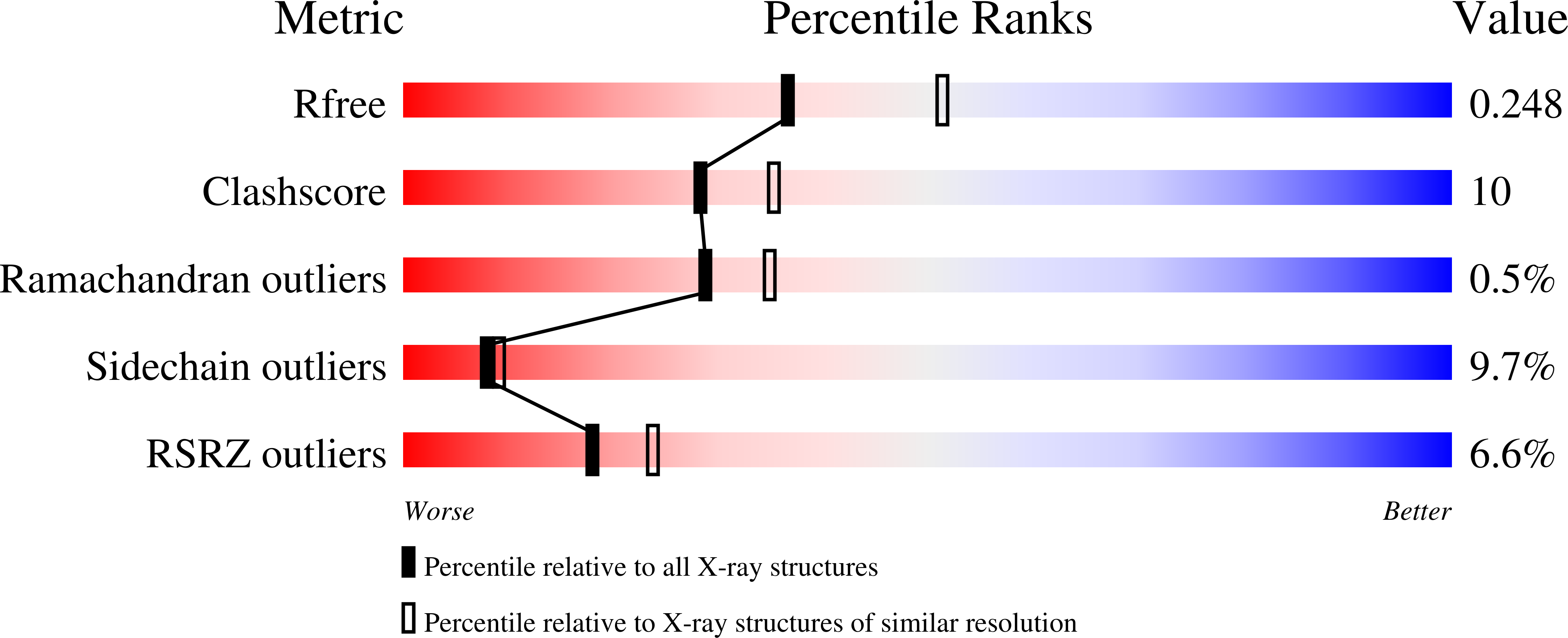
wwPDB Validation   3D Report Full Report


Ligand Structure Quality Assessment 


This is version 1.4 of the entry. See complete history


Literature

Coiled-Coil Helix Rotation Selects Repressing or Activating State of Transcriptional Regulator DhaR.

Shi, R.McDonald, L.Cygler, M.Ekiel, I.

(2014) Structure 22: 478-487

  • DOI: https://doi.org/10.1016/j.str.2013.11.012
  • Primary Citation of Related Structures:  
    4LRX, 4LRY, 4LRZ

  • PubMed Abstract: 

    Escherichia coli dihydroxyacetone (Dha) kinase consists of two subunits, DhaK and DhaL. Transcription of dha operon is regulated by the DhaR transcription factor and its action is under control of the kinase subunits. DhaR is activated by interaction with DhaL while it is repressed by DhaK. We have determined the structures of DhaK and DhaL bound to the tandem GAF-like and PAS domains of the DhaR, providing an architectural model for how GAF/PAS tandem domains work together in binding protein partners. The structures reveal a mechanism of opposite transcriptional regulation by the DhaK and DhaL subunits. The kinase subunits interface with DhaR through surfaces that partially overlap with their active sites, allowing sensing of ATP- versus ADP-loaded DhaL subunit and also precluding a ternary complex between DhaK-DhaL and DhaR. The rotation of helices within the DhaR coiled-coil linker upon DhaL binding provides the mechanism for transmitting the binding signal from the GAF/PAS domains to the C-terminal DNA-binding domain.


  • Organizational Affiliation

    Département de Biochimie, de Microbiologie et de Bio-informatique, PROTEO, Université Laval, Pavillon Charles-Eugene-Marchand, Québec City, QC G1V 0A6, Canada; Institut de Biologie Intégrative et des Systèmes (IBIS), Université Laval, Pavillon Charles-Eugene-Marchand, Québec City, QC G1V 0A6, Canada.


Macromolecules
Find similar proteins by:  (by identity cutoff)  |  3D Structure
Entity ID: 1
MoleculeChains Sequence LengthOrganismDetailsImage
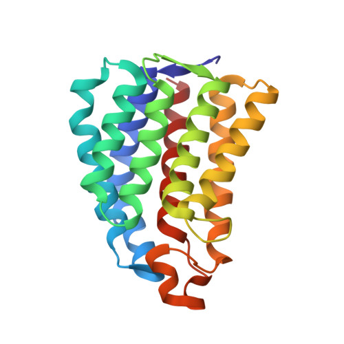
PTS-dependent dihydroxyacetone kinase, ADP-binding subunit DhaL
A, B, C, D
211Escherichia coli K-12Mutation(s): 0 
Gene Names: b1199dhaLdhaRJW5186ycgS
EC: 2.7 (PDB Primary Data), 2.7.1.121 (UniProt)
UniProt
Find proteins for P76014 (Escherichia coli (strain K12))
Explore P76014 
Go to UniProtKB:  P76014
Entity Groups  
Sequence Clusters30% Identity50% Identity70% Identity90% Identity95% Identity100% Identity
UniProt GroupP76014
Sequence Annotations
Expand
  • Reference Sequence
Find similar proteins by:  (by identity cutoff)  |  3D Structure
Entity ID: 2
MoleculeChains Sequence LengthOrganismDetailsImage
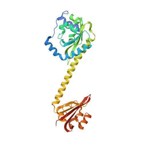
PTS-dependent dihydroxyacetone kinase operon regulatory protein
E, F, G, H
318Escherichia coli K-12Mutation(s): 0 
Gene Names: b1201dhaLdhaRJW5188ycgU
UniProt
Find proteins for P76016 (Escherichia coli (strain K12))
Explore P76016 
Go to UniProtKB:  P76016
Entity Groups  
Sequence Clusters30% Identity50% Identity70% Identity90% Identity95% Identity100% Identity
UniProt GroupP76016
Sequence Annotations
Expand
  • Reference Sequence
Small Molecules
Ligands 2 Unique
IDChains Name / Formula / InChI Key2D Diagram3D Interactions
ADP
Query on ADP

Download Ideal Coordinates CCD File 
I [auth A],
L [auth B],
O [auth C],
R [auth D]
ADENOSINE-5'-DIPHOSPHATE
C10 H15 N5 O10 P2
XTWYTFMLZFPYCI-KQYNXXCUSA-N
MG
Query on MG

Download Ideal Coordinates CCD File 
J [auth A]
K [auth A]
M [auth B]
N [auth B]
P [auth C]
J [auth A],
K [auth A],
M [auth B],
N [auth B],
P [auth C],
Q [auth C],
S [auth D],
T [auth D]
MAGNESIUM ION
Mg
JLVVSXFLKOJNIY-UHFFFAOYSA-N
Experimental Data & Validation

Experimental Data

  • Method: X-RAY DIFFRACTION
  • Resolution: 2.32 Å
  • R-Value Free: 0.247 
  • R-Value Work: 0.200 
  • R-Value Observed: 0.202 
  • Space Group: P 1
Unit Cell:
Length ( Å )Angle ( ˚ )
a = 89.766α = 84.15
b = 91.5β = 72.42
c = 93.808γ = 90.01
Software Package:
Software NamePurpose
DENZOdata reduction
SCALEPACKdata scaling
MOLREPphasing
REFMACrefinement
PDB_EXTRACTdata extraction
HKL-2000data collection
HKL-2000data reduction

Structure Validation

View Full Validation Report


Ligand Structure Quality Assessment 


Entry History 

Deposition Data

Revision History  (Full details and data files)

  • Version 1.0: 2014-01-29
    Type: Initial release
  • Version 1.1: 2014-02-05
    Changes: Data collection
  • Version 1.2: 2014-03-26
    Changes: Database references
  • Version 1.3: 2017-11-15
    Changes: Refinement description
  • Version 1.4: 2024-02-28
    Changes: Data collection, Database references, Derived calculations