


Target-directed self-assembly of homodimeric drugs
Giardina, S.F., Pingle, M., Foreman, K.W., Werner, D.S., Bergstrom, D.E., Barany, F., Arnold, L.D.To be published.
Experimental Data Snapshot
Entity ID: 1 | |||||
|---|---|---|---|---|---|
| Molecule | Chains | Sequence Length | Organism | Details | Image |
| Tryptase alpha/beta-1 | 245 | Homo sapiens | Mutation(s): 0 Gene Names: TPS1, TPS2, TPSAB1, TPSB1 EC: 3.4.21.59 | 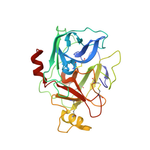 | |
UniProt & NIH Common Fund Data Resources | |||||
Find proteins for Q15661 (Homo sapiens) Explore Q15661 Go to UniProtKB: Q15661 | |||||
PHAROS: Q15661 GTEx: ENSG00000172236 | |||||
Entity Groups | |||||
| Sequence Clusters | 30% Identity50% Identity70% Identity90% Identity95% Identity100% Identity | ||||
| UniProt Group | Q15661 | ||||
Sequence AnnotationsExpand | |||||
| |||||
| Ligands 3 Unique | |||||
|---|---|---|---|---|---|
| ID | Chains | Name / Formula / InChI Key | 2D Diagram | 3D Interactions | |
| X00 Query on X00 | F [auth B] | {(1,1,3,3-tetramethyldisiloxane-1,3-diyl)bis[5-(methylsulfanyl)benzene-3,1-diyl]}bis({4-[3-(aminomethyl)phenyl]piperidin-1-yl}methanone) C44 H58 N4 O3 S2 Si2 MOGPKFKDWOMWNP-UHFFFAOYSA-N |  | ||
| PG4 Query on PG4 | D [auth A] | TETRAETHYLENE GLYCOL C8 H18 O5 UWHCKJMYHZGTIT-UHFFFAOYSA-N |  | ||
| SO4 Query on SO4 | C [auth A], E [auth A], G [auth B], H [auth B], I [auth B] | SULFATE ION O4 S QAOWNCQODCNURD-UHFFFAOYSA-L |  | ||
| Length ( Å ) | Angle ( ˚ ) |
|---|---|
| a = 78.168 | α = 90 |
| b = 78.168 | β = 90 |
| c = 165.458 | γ = 120 |
| Software Name | Purpose |
|---|---|
| SCALEPACK | data scaling |
| REFMAC | refinement |
| PDB_EXTRACT | data extraction |
| ADSC | data collection |
| HKL-2000 | data reduction |
| PHASER | phasing |


