4N1E

Structural evidence for antigen receptor evolution


Experimental Data Snapshot

  • Method: X-RAY DIFFRACTION
  • Resolution: 2.23 Å
  • R-Value Free: 0.265 
  • R-Value Work: 0.223 
  • R-Value Observed: 0.225 

wwPDB Validation   3D Report Full Report


This is version 1.2 of the entry. See complete history


Literature

Structural reconstruction of protein ancestry.

Rouet, R.Langley, D.B.Schofield, P.Christie, M.Roome, B.Porebski, B.T.Buckle, A.M.Clifton, B.E.Jackson, C.J.Stock, D.Christ, D.

(2017) Proc Natl Acad Sci U S A 114: 3897-3902

  • DOI: https://doi.org/10.1073/pnas.1613477114
  • Primary Citation of Related Structures:  
    4N1C, 4N1E

  • PubMed Abstract: 

    Ancestral protein reconstruction allows the resurrection and characterization of ancient proteins based on computational analyses of sequences of modern-day proteins. Unfortunately, many protein families are highly divergent and not suitable for sequence-based reconstruction approaches. This limitation is exemplified by the antigen receptors of jawed vertebrates (B- and T-cell receptors), heterodimers formed by pairs of Ig domains. These receptors are believed to have evolved from an extinct homodimeric ancestor through a process of gene duplication and diversification; however molecular evidence has so far remained elusive. Here, we use a structural approach and laboratory evolution to reconstruct such molecules and characterize their interaction with antigen. High-resolution crystal structures of reconstructed homodimeric receptors in complex with hen-egg white lysozyme demonstrate how nanomolar affinity binding of asymmetrical antigen is enabled through selective recruitment and structural plasticity within the receptor-binding site. Our results provide structural evidence in support of long-held theories concerning the evolution of antigen receptors, and provide a blueprint for the experimental reconstruction of protein ancestry in the absence of phylogenetic evidence.


  • Organizational Affiliation

    Garvan Institute of Medical Research, Darlinghurst, Sydney, NSW 2010, Australia.


Macromolecules
Find similar proteins by:  (by identity cutoff)  |  3D Structure
Entity ID: 1
MoleculeChains Sequence LengthOrganismDetailsImage
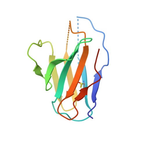
immunoglobulin variable light chain domain
A, B, C, D, E
A, B, C, D, E, F, G, H
109Homo sapiensMutation(s): 0 
Entity Groups  
Sequence Clusters30% Identity50% Identity70% Identity90% Identity95% Identity100% Identity
Sequence Annotations
Expand
  • Reference Sequence
Find similar proteins by:  (by identity cutoff)  |  3D Structure
Entity ID: 2
MoleculeChains Sequence LengthOrganismDetailsImage
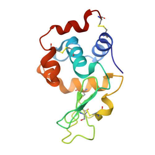
Lysozyme C
I, J, K, L
129Gallus gallusMutation(s): 0 
EC: 3.2.1.17
UniProt
Find proteins for P00698 (Gallus gallus)
Explore P00698 
Go to UniProtKB:  P00698
Entity Groups  
Sequence Clusters30% Identity50% Identity70% Identity90% Identity95% Identity100% Identity
UniProt GroupP00698
Sequence Annotations
Expand
  • Reference Sequence
Experimental Data & Validation

Experimental Data

  • Method: X-RAY DIFFRACTION
  • Resolution: 2.23 Å
  • R-Value Free: 0.265 
  • R-Value Work: 0.223 
  • R-Value Observed: 0.225 
  • Space Group: P 1 21 1
Unit Cell:
Length ( Å )Angle ( ˚ )
a = 114.917α = 90
b = 39.413β = 106.38
c = 178.301γ = 90
Software Package:
Software NamePurpose
MOSFLMdata reduction
SCALAdata scaling
PHASERphasing
REFMACrefinement
PDB_EXTRACTdata extraction
MAR345dtbdata collection

Structure Validation

View Full Validation Report


Entry History 

Deposition Data

Revision History  (Full details and data files)

  • Version 1.0: 2014-10-29
    Type: Initial release
  • Version 1.1: 2017-03-29
    Changes: Database references
  • Version 1.2: 2018-04-18
    Changes: Data collection, Database references