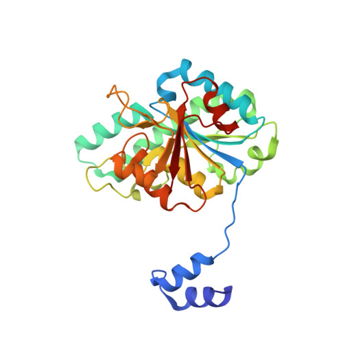
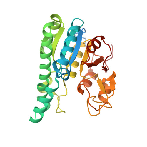
4NDR

Crystal structure Molybdenum Storage Protein with fully Mo-loaded cavity


Experimental Data Snapshot

  • Method: X-RAY DIFFRACTION
  • Resolution: 2.00 Å
  • R-Value Free: 0.191 
  • R-Value Work: 0.164 
  • R-Value Observed: 0.165 

Starting Model: experimental
View more details

wwPDB Validation   3D Report Full Report


Ligand Structure Quality Assessment 


This is version 1.4 of the entry. See complete history


Literature

Structural diversity of polyoxomolybdate clusters along the three-fold axis of the molybdenum storage protein.

Poppe, J.Warkentin, E.Demmer, U.Kowalewski, B.Dierks, T.Schneider, K.Ermler, U.

(2014) J Inorg Biochem 138: 122-128

  • DOI: https://doi.org/10.1016/j.jinorgbio.2014.05.009
  • Primary Citation of Related Structures:  
    4NDO, 4NDP, 4NDQ, 4NDR

  • PubMed Abstract: 

    The molybdenum storage protein (MoSto) can store more than 100 Mo or W atoms as discrete polyoxometalate (POM) clusters. Here, we describe the three POM cluster sites along the threefold axis of the protein complex based on four X-ray structures with slightly different polyoxomolybdate compositions between 1.35 and 2 Å resolution. In contrast to the Moα-out binding site occupied by an Mo3 cluster, the Moα-in and Moβ binding sites contain rather weak and non-uniform electron density for the Mo atoms (but clearly identifiable by anomalous data), suggesting the presence of POM cluster ensembles and/or degradation products of larger aggregates. The "Moα-in cluster ensemble" was interpreted as an antiprism-like Mo6 species superimposed with an Mo7 pyramide and the "Moβ cluster ensemble" as an Mo13 cluster (present mostly in a degraded form) composed of a pyramidal Mo7 and a Mo3 building block linked by three spatially separated MoOx units. Inside the ball-shaped Mo13 cluster sits an occluded central atom, perhaps a metal ion. POM cluster formation at the Moα-in and Moβ sites appears to be driven by filtering out and binding/protecting self-assembled transient species complementary to the protein template.


  • Organizational Affiliation

    Max-Planck-Institut für Biophysik, Max-von-Laue-Straße 3, D-60438 Frankfurt am Main, Germany.


Macromolecules
Find similar proteins by:  (by identity cutoff)  |  3D Structure
Entity ID: 1
MoleculeChains Sequence LengthOrganismDetailsImage
Molybdenum storage protein subunit betaA [auth B]270Azotobacter vinelandii DJMutation(s): 0 
UniProt
Find proteins for P84253 (Azotobacter vinelandii (strain DJ / ATCC BAA-1303))
Explore P84253 
Go to UniProtKB:  P84253
Entity Groups  
Sequence Clusters30% Identity50% Identity70% Identity90% Identity95% Identity100% Identity
UniProt GroupP84253
Sequence Annotations
Expand
  • Reference Sequence
Find similar proteins by:  (by identity cutoff)  |  3D Structure
Entity ID: 2
MoleculeChains Sequence LengthOrganismDetailsImage
Molybdenum storage protein subunit alphaB [auth A]276Azotobacter vinelandii DJMutation(s): 0 
UniProt
Find proteins for P84308 (Azotobacter vinelandii (strain DJ / ATCC BAA-1303))
Explore P84308 
Go to UniProtKB:  P84308
Entity Groups  
Sequence Clusters30% Identity50% Identity70% Identity90% Identity95% Identity100% Identity
UniProt GroupP84308
Sequence Annotations
Expand
  • Reference Sequence
Small Molecules
Ligands 6 Unique
IDChains Name / Formula / InChI Key2D Diagram3D Interactions
8M0
Query on 8M0

Download Ideal Coordinates CCD File 
E [auth B],
X [auth A]
bis(mu4-oxo)-tetrakis(mu3-oxo)-hexakis(mu2-oxo)-hexadecaoxo-octamolybdenum (VI)
Mo8 O28
GSOSAILZTJNYOK-UHFFFAOYSA-N
ATP
Query on ATP

Download Ideal Coordinates CCD File 
C [auth B],
V [auth A]
ADENOSINE-5'-TRIPHOSPHATE
C10 H16 N5 O13 P3
ZKHQWZAMYRWXGA-KQYNXXCUSA-N
M10
Query on M10

Download Ideal Coordinates CCD File 
Y [auth A](mu3-oxo)-tris(mu2-oxo)-nonakisoxo-trimolybdenum (VI)
Mo3 O13
HGNGIYFURFJYFJ-UHFFFAOYSA-N
MO
Query on MO

Download Ideal Coordinates CCD File 
AA [auth A]
BA [auth A]
CA [auth A]
F [auth B]
G [auth B]
AA [auth A],
BA [auth A],
CA [auth A],
F [auth B],
G [auth B],
H [auth B],
I [auth B],
J [auth B],
K [auth B],
L [auth B],
M [auth B],
N [auth B],
O [auth B],
P [auth B],
Q [auth B],
R [auth B],
S [auth B],
T [auth B],
U [auth B],
Z [auth A]
MOLYBDENUM ATOM
Mo
ZOKXTWBITQBERF-UHFFFAOYSA-N
PO4
Query on PO4

Download Ideal Coordinates CCD File 
D [auth B]PHOSPHATE ION
O4 P
NBIIXXVUZAFLBC-UHFFFAOYSA-K
MG
Query on MG

Download Ideal Coordinates CCD File 
W [auth A]MAGNESIUM ION
Mg
JLVVSXFLKOJNIY-UHFFFAOYSA-N
Experimental Data & Validation

Experimental Data

  • Method: X-RAY DIFFRACTION
  • Resolution: 2.00 Å
  • R-Value Free: 0.191 
  • R-Value Work: 0.164 
  • R-Value Observed: 0.165 
  • Space Group: P 63 2 2
Unit Cell:
Length ( Å )Angle ( ˚ )
a = 114.27α = 90
b = 114.27β = 90
c = 234.14γ = 120
Software Package:
Software NamePurpose
PHENIXrefinement
REFMACrefinement
XDSdata reduction
XDSdata scaling
REFMACphasing

Structure Validation

View Full Validation Report


Ligand Structure Quality Assessment 


Entry History 

Deposition Data

Revision History  (Full details and data files)

  • Version 1.0: 2014-08-13
    Type: Initial release
  • Version 1.1: 2014-11-12
    Changes: Structure summary
  • Version 1.2: 2017-11-15
    Changes: Refinement description
  • Version 1.3: 2019-07-17
    Changes: Data collection, Refinement description
  • Version 1.4: 2023-09-20
    Changes: Data collection, Database references, Derived calculations, Refinement description