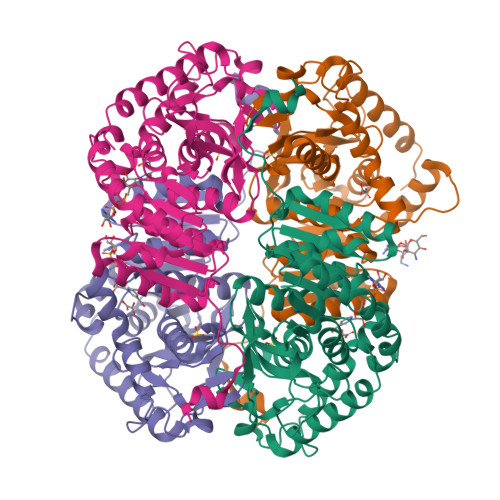
4OKN

Crystal structure of human muscle L-lactate dehydrogenase, ternary complex with NADH and oxalate


Experimental Data Snapshot

  • Method: X-RAY DIFFRACTION
  • Resolution: 2.10 Å
  • R-Value Free: 0.245 
  • R-Value Work: 0.206 
  • R-Value Observed: 0.208 

wwPDB Validation   3D Report Full Report


Ligand Structure Quality Assessment 


This is version 1.3 of the entry. See complete history


Literature

Structures of lactate dehydrogenase A (LDHA) in apo, ternary and inhibitor-bound forms.

Kolappan, S.Shen, D.L.Mosi, R.Sun, J.McEachern, E.J.Vocadlo, D.J.Craig, L.

(2015) Acta Crystallogr D Biol Crystallogr 71: 185-195

  • DOI: https://doi.org/10.1107/S1399004714024791
  • Primary Citation of Related Structures:  
    4OJN, 4OKN, 4QSM, 4QT0

  • PubMed Abstract: 

    Lactate dehydrogenase (LDH) is an essential metabolic enzyme that catalyzes the interconversion of pyruvate and lactate using NADH/NAD(+) as a co-substrate. Many cancer cells exhibit a glycolytic phenotype known as the Warburg effect, in which elevated LDH levels enhance the conversion of glucose to lactate, making LDH an attractive therapeutic target for oncology. Two known inhibitors of the human muscle LDH isoform, LDHA, designated 1 and 2, were selected, and their IC50 values were determined to be 14.4 ± 3.77 and 2.20 ± 0.15 µM, respectively. The X-ray crystal structures of LDHA in complex with each inhibitor were determined; both inhibitors bind to a site overlapping with the NADH-binding site. Further, an apo LDHA crystal structure solved in a new space group is reported, as well as a complex with both NADH and the substrate analogue oxalate bound in seven of the eight molecules and an oxalate only bound in the eighth molecule in the asymmetric unit. In this latter structure, a kanamycin molecule is located in the inhibitor-binding site, thereby blocking NADH binding. These structures provide insights into LDHA enzyme mechanism and inhibition and a framework for structure-assisted drug design that may contribute to new cancer therapies.


  • Organizational Affiliation

    Department of Molecular Biology and Biochemistry, Simon Fraser University, Burnaby, BC V5A 3Y6, Canada.


Macromolecules
Find similar proteins by:  (by identity cutoff)  |  3D Structure
Entity ID: 1
MoleculeChains Sequence LengthOrganismDetailsImage
L-lactate dehydrogenase A chain
A, B, C, D, E
A, B, C, D, E, F, G, H
337Homo sapiensMutation(s): 0 
Gene Names: LDHAPIG19
EC: 1.1.1.27
UniProt & NIH Common Fund Data Resources
Find proteins for P00338 (Homo sapiens)
Explore P00338 
Go to UniProtKB:  P00338
PHAROS:  P00338
GTEx:  ENSG00000134333 
Entity Groups  
Sequence Clusters30% Identity50% Identity70% Identity90% Identity95% Identity100% Identity
UniProt GroupP00338
Sequence Annotations
Expand
  • Reference Sequence
Small Molecules
Ligands 4 Unique
IDChains Name / Formula / InChI Key2D Diagram3D Interactions
NAI
Query on NAI

Download Ideal Coordinates CCD File 
BA [auth G]
EA [auth H]
I [auth A]
O [auth C]
R [auth D]
BA [auth G],
EA [auth H],
I [auth A],
O [auth C],
R [auth D],
U [auth E],
Y [auth F]
1,4-DIHYDRONICOTINAMIDE ADENINE DINUCLEOTIDE
C21 H29 N7 O14 P2
BOPGDPNILDQYTO-NNYOXOHSSA-N
KAN
Query on KAN

Download Ideal Coordinates CCD File 
N [auth B]KANAMYCIN A
C18 H36 N4 O11
SBUJHOSQTJFQJX-NOAMYHISSA-N
SO4
Query on SO4

Download Ideal Coordinates CCD File 
AA [auth F]
DA [auth G]
GA [auth H]
K [auth A]
M [auth B]
AA [auth F],
DA [auth G],
GA [auth H],
K [auth A],
M [auth B],
Q [auth C],
T [auth D],
W [auth E],
X [auth E]
SULFATE ION
O4 S
QAOWNCQODCNURD-UHFFFAOYSA-L
OXL
Query on OXL

Download Ideal Coordinates CCD File 
CA [auth G]
FA [auth H]
J [auth A]
L [auth B]
P [auth C]
CA [auth G],
FA [auth H],
J [auth A],
L [auth B],
P [auth C],
S [auth D],
V [auth E],
Z [auth F]
OXALATE ION
C2 O4
MUBZPKHOEPUJKR-UHFFFAOYSA-L
Experimental Data & Validation

Experimental Data

  • Method: X-RAY DIFFRACTION
  • Resolution: 2.10 Å
  • R-Value Free: 0.245 
  • R-Value Work: 0.206 
  • R-Value Observed: 0.208 
  • Space Group: P 21 21 21
Unit Cell:
Length ( Å )Angle ( ˚ )
a = 65.55α = 90
b = 159.62β = 90
c = 266.16γ = 90
Software Package:
Software NamePurpose
Blu-Icedata collection
PHASERphasing
REFMACrefinement
XDSdata reduction
XSCALEdata scaling

Structure Validation

View Full Validation Report


Ligand Structure Quality Assessment 


Entry History 

Deposition Data

Revision History  (Full details and data files)

  • Version 1.0: 2014-12-03
    Type: Initial release
  • Version 1.1: 2015-02-11
    Changes: Database references
  • Version 1.2: 2015-02-25
    Changes: Database references
  • Version 1.3: 2024-02-28
    Changes: Data collection, Database references, Derived calculations