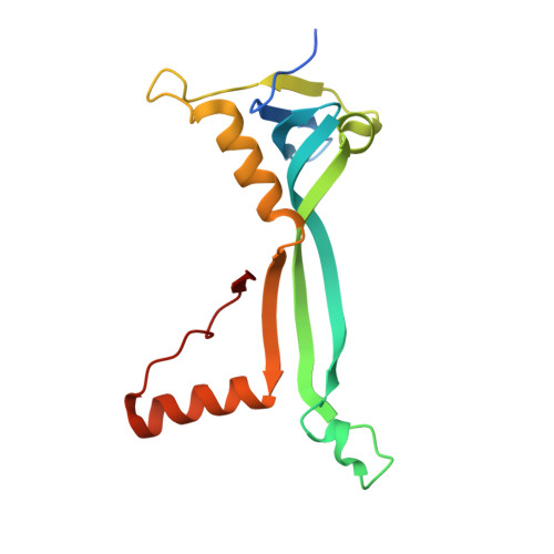
4Q95

Crystal structure of HRASLS3/LRAT chimeric protein


Experimental Data Snapshot

  • Method: X-RAY DIFFRACTION
  • Resolution: 2.20 Å
  • R-Value Free: 0.248 
  • R-Value Work: 0.207 
  • R-Value Observed: 0.209 

Starting Model: experimental
View more details

wwPDB Validation   3D Report Full Report


This is version 1.3 of the entry. See complete history


Literature

LRAT-specific domain facilitates vitamin A metabolism by domain swapping in HRASLS3.

Golczak, M.Sears, A.E.Kiser, P.D.Palczewski, K.

(2015) Nat Chem Biol 11: 26-32

  • DOI: https://doi.org/10.1038/nchembio.1687
  • Primary Citation of Related Structures:  
    4Q95

  • PubMed Abstract: 

    Cellular uptake of vitamin A, production of visual chromophore and triglyceride homeostasis in adipocytes depend on two representatives of the vertebrate N1pC/P60 protein family, lecithin:retinol acyltransferase (LRAT) and HRAS-like tumor suppressor 3 (HRASLS3). Both proteins function as lipid-metabolizing enzymes but differ in their substrate preferences and dominant catalytic activity. The mechanism of this catalytic diversity is not understood. Here, by using a gain-of-function approach, we identified a specific sequence responsible for the substrate specificity of N1pC/P60 proteins. A 2.2-Å crystal structure of the HRASLS3-LRAT chimeric enzyme in a thioester catalytic intermediate state revealed a major structural rearrangement accompanied by three-dimensional domain swapping dimerization not observed in native HRASLS proteins. Structural changes affecting the active site environment contributed to slower hydrolysis of the catalytic intermediate, supporting efficient acyl transfer. These findings reveal structural adaptation that facilitates selective catalysis and mechanism responsible for diverse substrate specificity within the LRAT-like enzyme family.


  • Organizational Affiliation

    Department of Pharmacology, Cleveland Center for Membrane and Structural Biology, School of Medicine, Case Western Reserve University, Cleveland, Ohio, USA.


Macromolecules
Find similar proteins by:  (by identity cutoff)  |  3D Structure
Entity ID: 1
MoleculeChains Sequence LengthOrganismDetailsImage
HRAS-like suppressor 3, Lecithin retinol acyltransferase
A, B
149Homo sapiensMus musculusMutation(s): 0 
Gene Names: HRASLS3HREV107human PLA2G16 and mouse LratLratPLA2G16
EC: 2.3.1.135
UniProt & NIH Common Fund Data Resources
Find proteins for P53816 (Homo sapiens)
Go to UniProtKB:  P53816
PHAROS:  P53816
GTEx:  ENSG00000176485 
Find proteins for Q9JI60 (Mus musculus)
Go to UniProtKB:  Q9JI60
IMPC:  MGI:1891259
Sequence Annotations
Expand
  • Reference Sequence
Small Molecules
Experimental Data & Validation

Experimental Data

  • Method: X-RAY DIFFRACTION
  • Resolution: 2.20 Å
  • R-Value Free: 0.248 
  • R-Value Work: 0.207 
  • R-Value Observed: 0.209 
  • Space Group: P 43 21 2
Unit Cell:
Length ( Å )Angle ( ˚ )
a = 63.123α = 90
b = 63.123β = 90
c = 157.965γ = 90
Software Package:
Software NamePurpose
XDSdata scaling
PHASERphasing
REFMACrefinement
XDSdata reduction

Structure Validation

View Full Validation Report


Entry History 

Deposition Data

Revision History  (Full details and data files)

  • Version 1.0: 2014-10-29
    Type: Initial release
  • Version 1.1: 2015-02-25
    Changes: Database references
  • Version 1.2: 2017-08-16
    Changes: Refinement description, Source and taxonomy
  • Version 1.3: 2023-09-20
    Changes: Data collection, Database references, Derived calculations, Refinement description