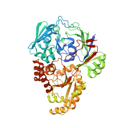
Structural insights into the multispecific recognition of dipeptides of deep-sea gram-negative bacterium Pseudoalteromonas sp. strain SM9913
Li, C.Y., Chen, X.L., Qin, Q.L., Wang, P., Zhang, W.X., Xie, B.B., Su, H.N., Zhang, X.Y., Zhou, B.C., Zhang, Y.Z.(2015) J Bacteriol 197: 1125-1134
- PubMed: 25605306
- DOI: https://doi.org/10.1128/JB.02600-14
- Primary Citation of Related Structures:
4QFK, 4QFL, 4QFN, 4QFO, 4QFP - PubMed Abstract:
Peptide uptake is important for nutrition supply for marine bacteria. It is also an important step in marine nitrogen cycling. However, how marine bacteria absorb peptides is still not fully understood. DppA is the periplasmic dipeptide binding protein of dipeptide permease (Dpp; an important peptide transporter in bacteria) and exclusively controls the substrate specificity of Dpp. Here, the substrate binding specificity of deep-sea Pseudoalteromonas sp. strain SM9913 DppA (PsDppA) was analyzed for 25 different dipeptides with various properties by using isothermal titration calorimetry measurements. PsDppA showed binding affinities for 8 dipeptides. To explain the multispecific substrate recognition mechanism of PsDppA, we solved the crystal structures of unliganded PsDppA and of PsDppA in complex with 4 different types of dipeptides (Ala-Phe, Met-Leu, Gly-Glu, and Val-Thr). PsDppA alternates between an "open" and a "closed" form during substrate binding. Structural analyses of the 4 PsDppA-substrate complexes combined with mutational assays indicate that PsDppA binds to different substrates through a precise mechanism: dipeptides are bound mainly by the interactions between their backbones and PsDppA, in particular by anchoring their N and C termini through ion-pair interactions; hydrophobic interactions are important in binding hydrophobic dipeptides; and Lys457 is necessary for the binding of dipeptides with a C-terminal glutamic acid or glutamine. Additionally, sequence alignment suggests that the substrate recognition mechanism of PsDppA may be common in Gram-negative bacteria. All together, our results provide structural insights into the multispecific substrate recognition mechanism of marine Gram-negative bacterial DppA, which provides a better understanding of the mechanisms of marine bacterial peptide uptake. Peptide uptake plays a significant role in nutrition supply for marine bacteria. It is also an important step in marine nitrogen cycling. However, how marine bacteria recognize and absorb peptides is still unclear. This study analyzed the substrate binding specificity of deep-sea Pseudoalteromonas sp. strain SM9913 DppA (PsDppA; the dipeptide-binding protein of dipeptide permease) and solved the crystal structures of unliganded PsDppA and PsDppA in complex with 4 different types of dipeptides. The multispecific recognition mechanism of PsDppA for dipeptides is explained based on structural and mutational analyses. We also find that the substrate-binding mechanism of PsDppA may be common in Gram-negative bacteria. This study sheds light on marine Gram-negative bacterial peptide uptake and marine nitrogen cycling.
Organizational Affiliation:
State Key Laboratory of Microbial Technology, Shandong University, Jinan, China Marine Biotechnology Research Center, Shandong University, Jinan, China.


