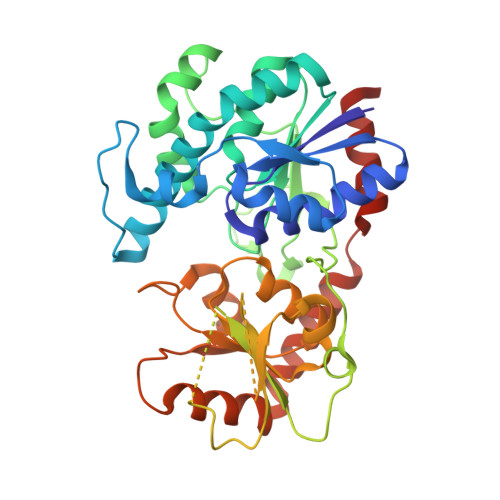
4RIG | pdb_00004rig

Chimeric Glycosyltransferase LanGT2S8Ac


Experimental Data Snapshot

  • Method: X-RAY DIFFRACTION
  • Resolution: 1.90 Å
  • R-Value Free: 
    0.241 (Depositor), 0.240 (DCC) 
  • R-Value Work: 
    0.203 (Depositor), 0.202 (DCC) 
  • R-Value Observed: 
    0.205 (Depositor) 

Starting Model: experimental
View more details

wwPDB Validation 3D Report Full Report

Validation slider image for 4RIG

This is version 1.5 of the entry. See complete history. 

Literature

Structural Characterization of O- and C-Glycosylating Variants of the Landomycin Glycosyltransferase LanGT2.

Tam, H.K., Harle, J., Gerhardt, S., Rohr, J., Wang, G., Thorson, J.S., Bigot, A., Lutterbeck, M., Seiche, W., Breit, B., Bechthold, A., Einsle, O.

(2015) Angew Chem Int Ed Engl 54: 2811-2815

  • DOI: https://doi.org/10.1002/anie.201409792
  • Primary Citation Related Structures: 
    4RIE, 4RIF, 4RIG, 4RIH, 4RII

  • PubMed Abstract: 

    The structures of the O-glycosyltransferase LanGT2 and the engineered, C-C bond-forming variant LanGT2S8Ac show how the replacement of a single loop can change the functionality of the enzyme. Crystal structures of the enzymes in complex with a nonhydrolyzable nucleotide-sugar analogue revealed that there is a conformational transition to create the binding sites for the aglycon substrate. This induced-fit transition was explored by molecular docking experiments with various aglycon substrates.


  • Organizational Affiliation: 
    • Institut für Biochemie, Albert-Ludwigs-Universität Freiburg, Albertstrasse 21, 79104 Freiburg (Germany).

Macromolecule Content 

  • Total Structure Weight: 80.95 kDa 
  • Atom Count: 5,450 
  • Modeled Residue Count: 703 
  • Deposited Residue Count: 758 
  • Unique protein chains: 1

Macromolecules

Find similar proteins by:|  3D Structure
Entity ID: 1
MoleculeChains  Sequence LengthOrganismDetailsImage
Glycosyl transferase
A, B
379Streptomyces cyanogenus, Streptomyces fradiaeMutation(s): 1 
Gene Names: lanGT2, urdGT2
UniProt
Find proteins for Q9RPA7 (Streptomyces fradiae)
Explore Q9RPA7 
Go to UniProtKB:  Q9RPA7
Find proteins for Q9ZGC0 (Streptomyces cyanogenus)
Explore Q9ZGC0 
Go to UniProtKB:  Q9ZGC0
Entity Groups
Sequence Clusters30% Identity50% Identity70% Identity90% Identity95% Identity100% Identity
UniProt GroupsQ9RPA7Q9ZGC0
Sequence Annotations
Expand
Reference Sequence

Experimental Data & Validation

Experimental Data

  • Method: X-RAY DIFFRACTION
  • Resolution: 1.90 Å
  • R-Value Free:  0.241 (Depositor), 0.240 (DCC) 
  • R-Value Work:  0.203 (Depositor), 0.202 (DCC) 
  • R-Value Observed: 0.205 (Depositor) 
Space Group: P 1
Unit Cell:
Length ( Å )Angle ( ˚ )
a = 52.943α = 79.3
b = 59.109β = 71.72
c = 63.111γ = 87.69
Software Package:
Software NamePurpose
MOLREPphasing
REFMACrefinement
XDSdata reduction
SCALAdata scaling

Structure Validation

View Full Validation Report


Entry History 

Deposition Data

Revision History  (Full details and data files)

  • Version 1.0: 2015-01-28
    Type: Initial release
  • Version 1.1: 2015-03-04
    Changes: Database references
  • Version 1.2: 2017-08-02
    Changes: Refinement description, Source and taxonomy
  • Version 1.3: 2017-11-22
    Changes: Refinement description
  • Version 1.4: 2024-02-28
    Changes: Data collection, Database references, Derived calculations, Refinement description
  • Version 1.5: 2024-04-03
    Changes: Refinement description