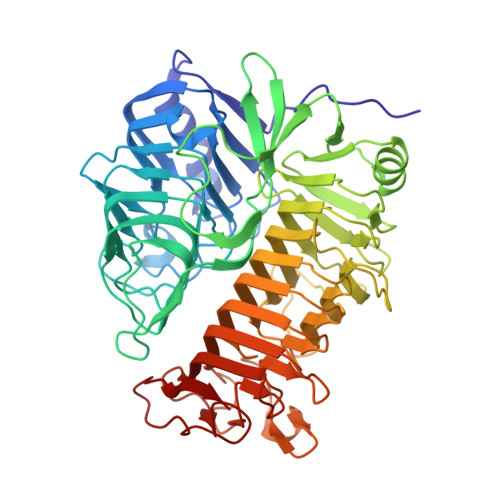
4TZ3

Ensemble refinement of the E502A variant of sacteLam55A from Streptomyces sp. SirexAA-E in complex with laminaritetraose


Experimental Data Snapshot

  • Method: X-RAY DIFFRACTION
  • Resolution: 1.90 Å
  • R-Value Free: 0.151 
  • R-Value Work: 0.113 
  • R-Value Observed: 0.115 

Starting Model: experimental
View more details

wwPDB Validation   3D Report Full Report


Ligand Structure Quality Assessment 


This is version 2.1 of the entry. See complete history

Re-refinement Note

This entry reflects an alternative modeling of the original data in: 4PEZ


Literature

Active site and laminarin binding in glycoside hydrolase family 55.

Bianchetti, C.M.Takasuka, T.E.Deutsch, S.Udell, H.S.Yik, E.J.Bergeman, L.F.Fox, B.G.

(2015) J Biol Chem 290: 11819-11832

  • DOI: https://doi.org/10.1074/jbc.M114.623579
  • Primary Citation of Related Structures:  
    4PEW, 4PEX, 4PEY, 4PEZ, 4PF0, 4TYV, 4TZ1, 4TZ3, 4TZ5

  • PubMed Abstract: 

    The Carbohydrate Active Enzyme (CAZy) database indicates that glycoside hydrolase family 55 (GH55) contains both endo- and exo-β-1,3-glucanases. The founding structure in the GH55 is PcLam55A from the white rot fungus Phanerochaete chrysosporium (Ishida, T., Fushinobu, S., Kawai, R., Kitaoka, M., Igarashi, K., and Samejima, M. (2009) Crystal structure of glycoside hydrolase family 55 β-1,3-glucanase from the basidiomycete Phanerochaete chrysosporium. J. Biol. Chem. 284, 10100-10109). Here, we present high resolution crystal structures of bacterial SacteLam55A from the highly cellulolytic Streptomyces sp. SirexAA-E with bound substrates and product. These structures, along with mutagenesis and kinetic studies, implicate Glu-502 as the catalytic acid (as proposed earlier for Glu-663 in PcLam55A) and a proton relay network of four residues in activating water as the nucleophile. Further, a set of conserved aromatic residues that define the active site apparently enforce an exo-glucanase reactivity as demonstrated by exhaustive hydrolysis reactions with purified laminarioligosaccharides. Two additional aromatic residues that line the substrate-binding channel show substrate-dependent conformational flexibility that may promote processive reactivity of the bound oligosaccharide in the bacterial enzymes. Gene synthesis carried out on ∼30% of the GH55 family gave 34 active enzymes (19% functional coverage of the nonredundant members of GH55). These active enzymes reacted with only laminarin from a panel of 10 different soluble and insoluble polysaccharides and displayed a broad range of specific activities and optima for pH and temperature. Application of this experimental method provides a new, systematic way to annotate glycoside hydrolase phylogenetic space for functional properties.


  • Organizational Affiliation

    From the Great Lakes Bioenergy Research Center, University of Wisconsin-Madison, Madison, Wisconsin 53706, the Department of Chemistry, University of Wisconsin-Oshkosh, Oshkosh, Wisconsin 54901.


Macromolecules
Find similar proteins by:  (by identity cutoff)  |  3D Structure
Entity ID: 1
MoleculeChains Sequence LengthOrganismDetailsImage
Putative secreted protein549Streptomyces sp. SirexAA-EMutation(s): 1 
Gene Names: SACTE_4363
EC: 3.2.1.58
UniProt
Find proteins for G2NFJ9 (Streptomyces sp. (strain SirexAA-E / ActE))
Explore G2NFJ9 
Go to UniProtKB:  G2NFJ9
Entity Groups  
Sequence Clusters30% Identity50% Identity70% Identity90% Identity95% Identity100% Identity
UniProt GroupG2NFJ9
Sequence Annotations
Expand
  • Reference Sequence
Oligosaccharides

Help

Entity ID: 2
MoleculeChains Length2D Diagram Glycosylation3D Interactions
beta-D-glucopyranose-(1-3)-beta-D-glucopyranose-(1-3)-beta-D-glucopyranose-(1-3)-beta-D-glucopyranose
B
4N/A
Glycosylation Resources
GlyTouCan:  G87841AX
GlyCosmos:  G87841AX
GlyGen:  G87841AX
Experimental Data & Validation

Experimental Data

  • Method: X-RAY DIFFRACTION
  • Resolution: 1.90 Å
  • R-Value Free: 0.151 
  • R-Value Work: 0.113 
  • R-Value Observed: 0.115 
  • Space Group: P 1 21 1
Unit Cell:
Length ( Å )Angle ( ˚ )
a = 51.276α = 90
b = 100.21β = 99.46
c = 54.223γ = 90
Software Package:
Software NamePurpose
PDB_EXTRACTdata extraction
PHASERphasing
PHENIXrefinement
DENZOdata reduction
SCALEPACKdata scaling

Structure Validation

View Full Validation Report


Ligand Structure Quality Assessment 


Entry History & Funding Information

Deposition Data


Funding OrganizationLocationGrant Number
Department of Energy (DOE, United States)United StatesDE-FC02-07ER64494

Revision History  (Full details and data files)

  • Version 1.0: 2015-03-18
    Type: Initial release
  • Version 1.1: 2015-03-25
    Changes: Atomic model, Database references, Derived calculations, Non-polymer description
  • Version 1.2: 2015-05-20
    Changes: Database references
  • Version 1.3: 2017-09-27
    Changes: Advisory, Author supporting evidence, Database references, Derived calculations, Refinement description, Source and taxonomy, Structure summary
  • Version 1.4: 2019-12-04
    Changes: Author supporting evidence, Data collection
  • Version 2.0: 2020-07-29
    Type: Remediation
    Reason: Carbohydrate remediation
    Changes: Advisory, Atomic model, Data collection, Derived calculations, Structure summary
  • Version 2.1: 2023-09-27
    Changes: Data collection, Database references, Derived calculations, Refinement description, Structure summary