4U2R | pdb_00004u2r

Crystal structure of the GLUR2 ligand binding core (S1S2J, flip variant) in the apo state


Experimental Data Snapshot

  • Method: X-RAY DIFFRACTION
  • Resolution: 1.41 Å
  • R-Value Free: 
    0.232 (Depositor), 0.230 (DCC) 
  • R-Value Work: 
    0.212 (Depositor), 0.209 (DCC) 
  • R-Value Observed: 
    0.213 (Depositor) 

wwPDB Validation 3D Report Full Report

Validation slider image for 4U2R

This is version 1.5 of the entry. See complete history

Literature

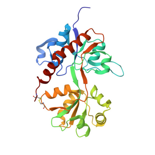
Structure and Dynamics of AMPA Receptor GluA2 in Resting, Pre-Open, and Desensitized States.

Durr, K.L.Chen, L.Stein, R.A.De Zorzi, R.Folea, I.M.Walz, T.Mchaourab, H.S.Gouaux, E.

(2014) Cell 158: 778-792

  • DOI: https://doi.org/10.1016/j.cell.2014.07.023
  • Primary Citation Related Structures: 
    4U1O, 4U1W, 4U1X, 4U1Y, 4U1Z, 4U21, 4U22, 4U23, 4U2P, 4U2Q, 4U2R

  • PubMed Abstract: 

    Ionotropic glutamate receptors (iGluRs) mediate the majority of fast excitatory signaling in the nervous system. Despite the profound importance of iGluRs to neurotransmission, little is known about the structures and dynamics of intact receptors in distinct functional states. Here, we elucidate the structures of the intact GluA2 AMPA receptor in an apo resting/closed state, in an activated/pre-open state bound with partial agonists and a positive allosteric modulator, and in a desensitized/closed state in complex with fluorowilliardiine. To probe the conformational properties of these states, we carried out double electron-electron resonance experiments on cysteine mutants and cryoelectron microscopy studies. We show how agonist binding modulates the conformation of the ligand-binding domain "layer" of the intact receptors and how, upon desensitization, the receptor undergoes large conformational rearrangements of the amino-terminal and ligand-binding domains. We define mechanistic principles by which to understand antagonism, activation, and desensitization in AMPA iGluRs.


  • Organizational Affiliation
    • Vollum Institute, Oregon Health and Science University, 3181 SW Sam Jackson Park Road, Portland, OR 97239, USA.

Macromolecule Content 

  • Total Structure Weight: 116.97 kDa 
  • Atom Count: 10,031 
  • Modeled Residue Count: 1,043 
  • Deposited Residue Count: 1,052 
  • Unique protein chains: 1

Macromolecules

Find similar proteins by:|  3D Structure
Entity ID: 1
MoleculeChains  Sequence LengthOrganismDetailsImage
Glutamate receptor 2A [auth B],
B [auth A],
C,
D
263Rattus norvegicusMutation(s): 1 
Gene Names: Gria2Glur2
UniProt
Find proteins for P19491 (Rattus norvegicus)
Explore P19491 
Go to UniProtKB:  P19491
Entity Groups
Sequence Clusters30% Identity50% Identity70% Identity90% Identity95% Identity100% Identity
UniProt GroupP19491
Sequence Annotations
Expand
Reference Sequence

Experimental Data & Validation

Experimental Data

  • Method: X-RAY DIFFRACTION
  • Resolution: 1.41 Å
  • R-Value Free:  0.232 (Depositor), 0.230 (DCC) 
  • R-Value Work:  0.212 (Depositor), 0.209 (DCC) 
  • R-Value Observed: 0.213 (Depositor) 
Space Group: P 21 21 21
Unit Cell:
Length ( Å )Angle ( ˚ )
a = 66.659α = 90
b = 91.936β = 90
c = 201.344γ = 90
Software Package:
Software NamePurpose
XDSdata reduction
PDB_EXTRACTdata extraction
PHENIXrefinement
PHASERphasing
Aimlessdata scaling

Structure Validation

View Full Validation Report


Entry History 

Deposition Data

Revision History  (Full details and data files)

  • Version 1.0: 2014-08-20
    Type: Initial release
  • Version 1.1: 2014-10-01
    Changes: Database references
  • Version 1.2: 2015-02-04
    Changes: Derived calculations
  • Version 1.3: 2017-11-22
    Changes: Advisory, Derived calculations, Other, Refinement description, Source and taxonomy
  • Version 1.4: 2023-12-27
    Changes: Data collection, Database references, Refinement description
  • Version 1.5: 2024-11-06
    Changes: Structure summary