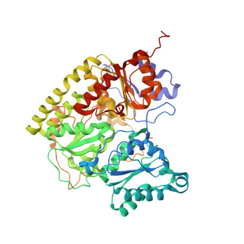
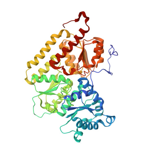
4WES

Nitrogenase molybdenum-iron protein from Clostridium pasteurianum at 1.08 A resolution


Experimental Data Snapshot

  • Method: X-RAY DIFFRACTION
  • Resolution: 1.08 Å
  • R-Value Free: 0.133 
  • R-Value Work: 0.110 
  • R-Value Observed: 0.111 

wwPDB Validation   3D Report Full Report


Ligand Structure Quality Assessment 


This is version 1.4 of the entry. See complete history


Literature

Nitrogenase MoFe protein from Clostridium pasteurianum at 1.08 angstrom resolution: comparison with the Azotobacter vinelandii MoFe protein.

Zhang, L.M.Morrison, C.N.Kaiser, J.T.Rees, D.C.

(2015) Acta Crystallogr D Biol Crystallogr 71: 274-282

  • DOI: https://doi.org/10.1107/S1399004714025243
  • Primary Citation of Related Structures:  
    4WES

  • PubMed Abstract: 

    The X-ray crystal structure of the nitrogenase MoFe protein from Clostridium pasteurianum (Cp1) has been determined at 1.08 Å resolution by multiwavelength anomalous diffraction phasing. Cp1 and the ortholog from Azotobacter vinelandii (Av1) represent two distinct families of nitrogenases, differing primarily by a long insertion in the α-subunit and a deletion in the β-subunit of Cp1 relative to Av1. Comparison of these two MoFe protein structures at atomic resolution reveals conserved structural arrangements that are significant to the function of nitrogenase. The FeMo cofactors defining the active sites of the MoFe protein are essentially identical between the two proteins. The surrounding environment is also highly conserved, suggesting that this structural arrangement is crucial for nitrogen reduction. The P clusters are likewise similar, although the surrounding protein and solvent environment is less conserved relative to that of the FeMo cofactor. The P cluster and FeMo cofactor in Av1 and Cp1 are connected through a conserved water tunnel surrounded by similar secondary-structure elements. The long α-subunit insertion loop occludes the presumed Fe protein docking surface on Cp1 with few contacts to the remainder of the protein. This makes it plausible that this loop is repositioned to open up the Fe protein docking surface for complex formation.


  • Organizational Affiliation

    Division of Chemistry and Chemical Engineering, California Institute of Technology, Pasadena, CA 91125, USA.


Macromolecules
Find similar proteins by:  (by identity cutoff)  |  3D Structure
Entity ID: 1
MoleculeChains Sequence LengthOrganismDetailsImage
Nitrogenase molybdenum-iron protein alpha chain
A, C
534Clostridium pasteurianum DSM 525 = ATCC 6013Mutation(s): 0 
EC: 1.18.6.1
UniProt
Find proteins for P00467 (Clostridium pasteurianum)
Go to UniProtKB:  P00467
Sequence Annotations
Expand
  • Reference Sequence
Find similar proteins by:  (by identity cutoff)  |  3D Structure
Entity ID: 2
MoleculeChains Sequence LengthOrganismDetailsImage
Nitrogenase molybdenum-iron protein beta chain
B, D
458Clostridium pasteurianum DSM 525 = ATCC 6013Mutation(s): 0 
EC: 1.18.6.1
UniProt
Find proteins for P11347 (Clostridium pasteurianum)
Go to UniProtKB:  P11347
Sequence Annotations
Expand
  • Reference Sequence
Small Molecules
Ligands 5 Unique
IDChains Name / Formula / InChI Key2D Diagram3D Interactions
ICS
Query on ICS

Download Ideal Coordinates CCD File 
F [auth A],
L [auth C]
iron-sulfur-molybdenum cluster with interstitial carbon
C Fe7 Mo S9
DDQFAOMIVKLFON-UHFFFAOYSA-N
CLF
Query on CLF

Download Ideal Coordinates CCD File 
H [auth A],
M [auth C]
FE(8)-S(7) CLUSTER
Fe8 S7
JKVMXLBGZBULKV-UHFFFAOYSA-N
HCA
Query on HCA

Download Ideal Coordinates CCD File 
E [auth A],
K [auth C]
3-HYDROXY-3-CARBOXY-ADIPIC ACID
C7 H10 O7
XKJVEVRQMLKSMO-SSDOTTSWSA-N
MPD
Query on MPD

Download Ideal Coordinates CCD File 
G [auth A](4S)-2-METHYL-2,4-PENTANEDIOL
C6 H14 O2
SVTBMSDMJJWYQN-YFKPBYRVSA-N
FE2
Query on FE2

Download Ideal Coordinates CCD File 
I [auth B],
J [auth B]
FE (II) ION
Fe
CWYNVVGOOAEACU-UHFFFAOYSA-N
Experimental Data & Validation

Experimental Data

  • Method: X-RAY DIFFRACTION
  • Resolution: 1.08 Å
  • R-Value Free: 0.133 
  • R-Value Work: 0.110 
  • R-Value Observed: 0.111 
  • Space Group: P 1 21 1
Unit Cell:
Length ( Å )Angle ( ˚ )
a = 72.701α = 90
b = 170.581β = 91.63
c = 87.54γ = 90
Software Package:
Software NamePurpose
REFMACrefinement
SCALAdata scaling
PDB_EXTRACTdata extraction
XSCALEdata scaling

Structure Validation

View Full Validation Report


Ligand Structure Quality Assessment 


Entry History & Funding Information

Deposition Data


Funding OrganizationLocationGrant Number
National Institutes of Health/National Institute of General Medical Sciences (NIH/NIGMS)United StatesGM45162
Natural Sciences and Engineering Research Council (NSERC, Canada)Canada--
National Science Foundation (NSF, United States)United StatesDGE-1144469

Revision History  (Full details and data files)

  • Version 1.0: 2015-02-11
    Type: Initial release
  • Version 1.1: 2015-02-25
    Changes: Database references
  • Version 1.2: 2017-09-27
    Changes: Advisory, Author supporting evidence, Derived calculations, Other, Refinement description, Source and taxonomy
  • Version 1.3: 2019-11-27
    Changes: Author supporting evidence
  • Version 1.4: 2023-12-27
    Changes: Data collection, Database references, Derived calculations, Refinement description