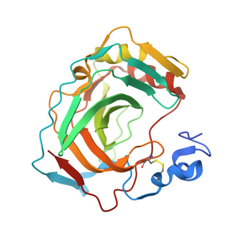
4X5S

The crystal structure of an alpha carbonic anhydrase from the extremophilic bacterium Sulfurihydrogenibium azorense.


Experimental Data Snapshot

  • Method: X-RAY DIFFRACTION
  • Resolution: 1.95 Å
  • R-Value Free: 0.214 
  • R-Value Work: 0.187 

Starting Model: experimental
View more details

wwPDB Validation   3D Report Full Report


Ligand Structure Quality Assessment 


This is version 1.1 of the entry. See complete history


Literature

Crystal structure of the most catalytically effective carbonic anhydrase enzyme known, SazCA from the thermophilic bacterium Sulfurihydrogenibium azorense.

De Simone, G.Monti, S.M.Alterio, V.Buonanno, M.De Luca, V.Rossi, M.Carginale, V.Supuran, C.T.Capasso, C.Di Fiore, A.

(2015) Bioorg Med Chem Lett 25: 2002-2006

  • DOI: https://doi.org/10.1016/j.bmcl.2015.02.068
  • Primary Citation of Related Structures:  
    4X5S

  • PubMed Abstract: 

    Two thermostable α-carbonic anhydrases (α-CAs) isolated from thermophilic Sulfurihydrogenibium spp., namely SspCA (from S. yellowstonensis) and SazCA (from S. azorense), were shown in a previous work to possess interesting complementary properties. SspCA was shown to have an exceptional thermal stability, whereas SazCA demonstrated to be the most active α-CA known to date for the CO2 hydration reaction. Here we report the crystallographic structure of SazCA and the identification of the structural features responsible for its high catalytic activity, by comparing it with SspCA structure. These data are of relevance for the design of engineered proteins showing higher stability and catalytic activity than other α-CAs known to date.


  • Organizational Affiliation

    Istituto di Biostrutture e Bioimmagini-CNR, via Mezzocannone 16, 80134 Napoli, Italy. Electronic address: gdesimon@unina.it.


Macromolecules
Find similar proteins by:  (by identity cutoff)  |  3D Structure
Entity ID: 1
MoleculeChains Sequence LengthOrganismDetailsImage
Carbonic anhydrase (Carbonate dehydratase)
A, B
232Sulfurihydrogenibium azorenseMutation(s): 0 
Gene Names: SULAZ_0541
EC: 4.2.1.1
UniProt
Find proteins for C1DTU5 (Sulfurihydrogenibium azorense (strain Az-Fu1 / DSM 15241 / OCM 825))
Explore C1DTU5 
Go to UniProtKB:  C1DTU5
Entity Groups  
Sequence Clusters30% Identity50% Identity70% Identity90% Identity95% Identity100% Identity
UniProt GroupC1DTU5
Sequence Annotations
Expand
  • Reference Sequence
Small Molecules
Ligands 3 Unique
IDChains Name / Formula / InChI Key2D Diagram3D Interactions
PE8
Query on PE8

Download Ideal Coordinates CCD File 
E [auth A],
H [auth B]
3,6,9,12,15,18,21-HEPTAOXATRICOSANE-1,23-DIOL
C16 H34 O9
GLZWNFNQMJAZGY-UHFFFAOYSA-N
AZM
Query on AZM

Download Ideal Coordinates CCD File 
D [auth A],
G [auth B]
5-ACETAMIDO-1,3,4-THIADIAZOLE-2-SULFONAMIDE
C4 H6 N4 O3 S2
BZKPWHYZMXOIDC-UHFFFAOYSA-N
ZN
Query on ZN

Download Ideal Coordinates CCD File 
C [auth A],
F [auth B]
ZINC ION
Zn
PTFCDOFLOPIGGS-UHFFFAOYSA-N
Experimental Data & Validation

Experimental Data

  • Method: X-RAY DIFFRACTION
  • Resolution: 1.95 Å
  • R-Value Free: 0.214 
  • R-Value Work: 0.187 
  • Space Group: P 21 21 21
Unit Cell:
Length ( Å )Angle ( ˚ )
a = 46.47α = 90
b = 89.43β = 90
c = 114.82γ = 90
Software Package:
Software NamePurpose
CNSrefinement
PDB_EXTRACTdata extraction
HKL-2000data reduction
SCALEPACKdata scaling
AMoREphasing

Structure Validation

View Full Validation Report


Ligand Structure Quality Assessment 


Entry History 

Deposition Data

Revision History  (Full details and data files)

  • Version 1.0: 2015-05-20
    Type: Initial release
  • Version 1.1: 2024-01-10
    Changes: Data collection, Database references, Derived calculations, Refinement description