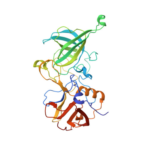
4Z4S

Crystal structure of GII.10 P domain in complex with 150mM fucose


Experimental Data Snapshot

  • Method: X-RAY DIFFRACTION
  • Resolution: 1.80 Å
  • R-Value Free: 0.182 
  • R-Value Work: 0.145 
  • R-Value Observed: 0.147 

Starting Model: experimental
View more details

wwPDB Validation   3D Report Full Report


Ligand Structure Quality Assessment 


This is version 1.3 of the entry. See complete history


Literature

The sweet quartet: Binding of fucose to the norovirus capsid.

Koromyslova, A.D.Leuthold, M.M.Bowler, M.W.Hansman, G.S.

(2015) Virology 483: 203-208

  • DOI: https://doi.org/10.1016/j.virol.2015.04.006
  • Primary Citation of Related Structures:  
    4Z4R, 4Z4S, 4Z4T, 4Z4U, 4Z4V, 4Z4W, 4Z4Y, 4Z4Z

  • PubMed Abstract: 

    Human noroviruses bind histo-blood group antigens (HBGAs) and this interaction is thought to be important for an infection. We identified two additional fucose-binding pockets (termed fucose-3/4 sites) on a genogroup II human (GII.10) norovirus-protruding (P) dimer using X-ray crystallography. Fucose-3/4 sites were located between two previously determined HBGA binding pockets (termed fucose-1/2 sites). We found that four fucose molecules were capable of binding altogether at fucose-1/2/3/4 sites on the P dimer, though the fucose molecules bound in a dose-dependent and step-wise manner. We also showed that HBGA B-trisaccharide molecules bound in a similar way at the fucose-1/2 sites. Interestingly, we discovered that the monomers of the P dimer were asymmetrical in an unliganded state and when a single B-trisaccharide molecule bound, but were symmetrical when two B-trisaccharide molecules bound. We postulate that the symmetrical dimers might favor HBGA binding interactions at fucose-1/2 sites.


  • Organizational Affiliation

    Schaller Research Group at the University of Heidelberg and the DKFZ, Heidelberg 69120, Germany; Department of Infectious Diseases, Virology, University of Heidelberg, Heidelberg 69120, Germany.


Macromolecules
Find similar proteins by:  (by identity cutoff)  |  3D Structure
Entity ID: 1
MoleculeChains Sequence LengthOrganismDetailsImage
Capsid protein
A, B
315Norovirus GII.10Mutation(s): 0 
UniProt
Find proteins for Q5F4T5 (Norwalk virus)
Explore Q5F4T5 
Go to UniProtKB:  Q5F4T5
Entity Groups  
Sequence Clusters30% Identity50% Identity70% Identity90% Identity95% Identity100% Identity
UniProt GroupQ5F4T5
Sequence Annotations
Expand
  • Reference Sequence
Small Molecules
Ligands 3 Unique
IDChains Name / Formula / InChI Key2D Diagram3D Interactions
FUL
Query on FUL

Download Ideal Coordinates CCD File 
G [auth A],
H [auth A],
M [auth B],
N [auth B]
beta-L-fucopyranose
C6 H12 O5
SHZGCJCMOBCMKK-KGJVWPDLSA-N
EDO
Query on EDO

Download Ideal Coordinates CCD File 
C [auth A]
D [auth A]
E [auth A]
F [auth A]
J [auth B]
C [auth A],
D [auth A],
E [auth A],
F [auth A],
J [auth B],
K [auth B],
L [auth B]
1,2-ETHANEDIOL
C2 H6 O2
LYCAIKOWRPUZTN-UHFFFAOYSA-N
NO3
Query on NO3

Download Ideal Coordinates CCD File 
I [auth A]NITRATE ION
N O3
NHNBFGGVMKEFGY-UHFFFAOYSA-N
Experimental Data & Validation

Experimental Data

  • Method: X-RAY DIFFRACTION
  • Resolution: 1.80 Å
  • R-Value Free: 0.182 
  • R-Value Work: 0.145 
  • R-Value Observed: 0.147 
  • Space Group: P 1 21 1
Unit Cell:
Length ( Å )Angle ( ˚ )
a = 66.09α = 90
b = 78.681β = 99.61
c = 68.811γ = 90
Software Package:
Software NamePurpose
PHASERphasing
PHENIXrefinement
PDB_EXTRACTdata extraction

Structure Validation

View Full Validation Report


Ligand Structure Quality Assessment 


Entry History 

Deposition Data

Revision History  (Full details and data files)

  • Version 1.0: 2015-05-27
    Type: Initial release
  • Version 1.1: 2015-06-03
    Changes: Database references
  • Version 1.2: 2020-07-29
    Type: Remediation
    Reason: Carbohydrate remediation
    Changes: Data collection, Derived calculations, Structure summary
  • Version 1.3: 2024-01-10
    Changes: Data collection, Database references, Refinement description, Structure summary