4ZHQ | pdb_00004zhq

Crystal structure of Tubulin-Stathmin-TTL-MMAE Complex


Experimental Data Snapshot

  • Method: X-RAY DIFFRACTION
  • Resolution: 2.55 Å
  • R-Value Free: 
    0.245 (Depositor), 0.246 (DCC) 
  • R-Value Work: 
    0.190 (Depositor), 0.192 (DCC) 
  • R-Value Observed: 
    0.192 (Depositor) 

Starting Model: experimental
View more details

wwPDB Validation 3D Report Full Report

Validation slider image for 4ZHQ

Ligand Structure Quality Assessment 


This is version 1.1 of the entry. See complete history

Literature

Structural Insights into the Pharmacophore of Vinca Domain Inhibitors of Microtubules

Wang, Y.Benz, F.W.Wu, Y.Wang, Q.Chen, Y.Chen, X.Li, H.Zhang, Y.Zhang, R.Yang, J.

(2016) Mol Pharmacol 89: 233-242

  • DOI: https://doi.org/10.1124/mol.115.100149
  • Primary Citation Related Structures: 
    4ZHQ, 4ZI7, 4ZOL, 5BMV

  • PubMed Abstract: 

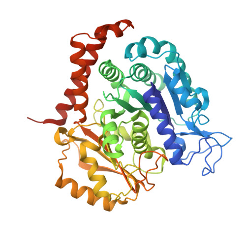
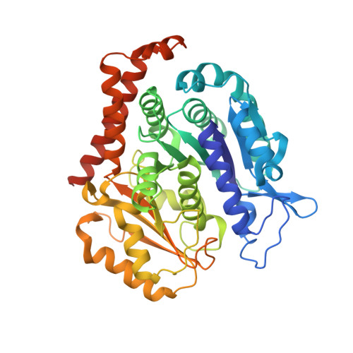
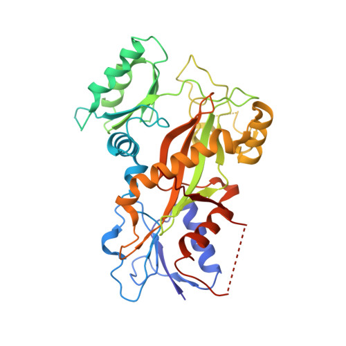
    Antibody-drug conjugates (ADCs) have achieved great success in cancer therapy in recent years. Some peptidyl microtubule inhibitors consisting of natural and unnatural amino acids, such as monomethyl auristatin E (MMAE) and F (MMAF), are extremely cytotoxic and have been used as a payload in ADCs. However, their precise molecular interaction with tubulin and microtubules remains unclear. We determined the crystal structures of tubulin in complex with three ultra-potent peptidyl microtubule inhibitors [MMAE, taltobulin (HTI- 286), and tubulysin M] at 2.5 Å. Our data showed that the three peptides bound to the vinca domain and shared a common and key pharmacophore containing two consecutive hydrophobic groups (Val, Ile-like side chain). These groups protruded in opposite directions into hydrophobic pockets on the tubulin β and α subunits. Nitrogen and oxygen atoms from the same backbone formed hydrogen bonds with Asn329 from the α subunit and Asp179 from the β subunit in a direction normal to the surface formed by the aforementioned hydrophobic groups. In addition, our crystal structure data indicated that tubulysin M bound to the β subunit alone, providing a structural explanation for its higher affinity. We also compared the conformations of two representative structurally different vinca domain compounds, ustiloxin D and vinblastine, with those of the aforementioned peptidyl ligands, and found that they shared a similar pharmacophore. Our findings lay a foundation for the rational design of novel vinca domain ligands and may facilitate the development of microtubule inhibitors with high specificity, affinity, and efficiency as payloads for ADCs in cancer therapy.


  • Organizational Affiliation
    • State Key Laboratory of Biotherapy and Cancer Center, West China Hospital, Sichuan University, and Collaborative Innovation Center for Biotherapy, Chengdu, China (Yu.W., Ya.W., Y.C., X.C., H.L., R.Z., J.Y.); Department of Pharmacology and Toxicology, University of Louisville School of Medicine, Health Sciences Center, Louisville, Kentucky (F.W.B.); Shanghai Institute of Applied Physics, Chinese Academy of Sciences, Shanghai, China (Q.W.); and Pharmacology and Pharmaceutical Sciences School of Medicine, Tsinghua University and Collaborative Innovation Center for Biotherapy, Beijing, China (Y.Z.).

Macromolecule Content 

  • Total Structure Weight: 265.95 kDa 
  • Atom Count: 17,988 
  • Modeled Residue Count: 2,197 
  • Deposited Residue Count: 2,319 
  • Unique protein chains: 4

Macromolecules

Find similar proteins by:|  3D Structure
Entity ID: 1
MoleculeChains  Sequence LengthOrganismDetailsImage
Tubulin alpha-1B chain
A, C
451Sus scrofaMutation(s): 0 
EC: 3.6.5
UniProt
Find proteins for Q2XVP4 (Sus scrofa)
Explore Q2XVP4 
Go to UniProtKB:  Q2XVP4
Entity Groups
Sequence Clusters30% Identity50% Identity70% Identity90% Identity95% Identity100% Identity
UniProt GroupQ2XVP4
Sequence Annotations
Expand
Reference Sequence
Find similar proteins by:|  3D Structure
Entity ID: 2
MoleculeChains  Sequence LengthOrganismDetailsImage
Tubulin beta chain
B, D
445Sus scrofaMutation(s): 0 
UniProt
Find proteins for P02554 (Sus scrofa)
Explore P02554 
Go to UniProtKB:  P02554
Entity Groups
Sequence Clusters30% Identity50% Identity70% Identity90% Identity95% Identity100% Identity
UniProt GroupP02554
Sequence Annotations
Expand
Reference Sequence
Find similar proteins by:|  3D Structure
Entity ID: 3
MoleculeChains  Sequence LengthOrganismDetailsImage
Stathmin-4143Rattus norvegicusMutation(s): 0 
Gene Names: Stmn4
UniProt
Find proteins for P63043 (Rattus norvegicus)
Explore P63043 
Go to UniProtKB:  P63043
Entity Groups
Sequence Clusters30% Identity50% Identity70% Identity90% Identity95% Identity100% Identity
UniProt GroupP63043
Sequence Annotations
Expand
Reference Sequence
Find similar proteins by:|  3D Structure
Entity ID: 4
MoleculeChains  Sequence LengthOrganismDetailsImage
Tubulin-Tyrosine Ligase384Gallus gallusMutation(s): 0 
Gene Names: TTL

Small Molecules

Ligands 8 Unique
IDChains Name / Formula / InChI Key2D Diagram3D Interactions
4Q5

Query on 4Q5


Download:Ideal Coordinates CCD File
S [auth B]N-methyl-L-valyl-N-[(3R,4S,5S)-1-{(2S)-2-[(1R,2R)-3-{[(1S,2R)-1-hydroxy-1-phenylpropan-2-yl]amino}-1-methoxy-2-methyl-3-oxopropyl]pyrrolidin-1-yl}-3-methoxy-5-methyl-1-oxoheptan-4-yl]-N-methyl-L-valinamide
C39 H67 N5 O7
DASWEROEPLKSEI-UIJRFTGLSA-N
GTP

Query on GTP


Download:Ideal Coordinates CCD File
G [auth A],
T [auth C]
GUANOSINE-5'-TRIPHOSPHATE
C10 H16 N5 O14 P3
XKMLYUALXHKNFT-UUOKFMHZSA-N
ACP

Query on ACP


Download:Ideal Coordinates CCD File
CA [auth F]PHOSPHOMETHYLPHOSPHONIC ACID ADENYLATE ESTER
C11 H18 N5 O12 P3
UFZTZBNSLXELAL-IOSLPCCCSA-N
GDP

Query on GDP


Download:Ideal Coordinates CCD File
AA [auth D],
M [auth B]
GUANOSINE-5'-DIPHOSPHATE
C10 H15 N5 O11 P2
QGWNDRXFNXRZMB-UUOKFMHZSA-N
MES

Query on MES


Download:Ideal Coordinates CCD File
P [auth B],
Q [auth B]
2-(N-MORPHOLINO)-ETHANESULFONIC ACID
C6 H13 N O4 S
SXGZJKUKBWWHRA-UHFFFAOYSA-N
GOL

Query on GOL


Download:Ideal Coordinates CCD File
J [auth A]
K [auth A]
L [auth A]
R [auth B]
V [auth C]
J [auth A],
K [auth A],
L [auth A],
R [auth B],
V [auth C],
X [auth C],
Y [auth C],
Z [auth C]
GLYCEROL
C3 H8 O3
PEDCQBHIVMGVHV-UHFFFAOYSA-N
CA

Query on CA


Download:Ideal Coordinates CCD File
I [auth A],
O [auth B],
W [auth C]
CALCIUM ION
Ca
BHPQYMZQTOCNFJ-UHFFFAOYSA-N
MG

Query on MG


Download:Ideal Coordinates CCD File
BA [auth D],
H [auth A],
N [auth B],
U [auth C]
MAGNESIUM ION
Mg
JLVVSXFLKOJNIY-UHFFFAOYSA-N

Experimental Data & Validation

Experimental Data

  • Method: X-RAY DIFFRACTION
  • Resolution: 2.55 Å
  • R-Value Free:  0.245 (Depositor), 0.246 (DCC) 
  • R-Value Work:  0.190 (Depositor), 0.192 (DCC) 
  • R-Value Observed: 0.192 (Depositor) 
Space Group: P 21 21 21
Unit Cell:
Length ( Å )Angle ( ˚ )
a = 104.938α = 90
b = 156.809β = 90
c = 182.465γ = 90
Software Package:
Software NamePurpose
REFMACrefinement
PDB_EXTRACTdata extraction
SCALEPACKdata reduction
SCALEPACKdata scaling
MLPHAREphasing

Structure Validation

View Full Validation Report


Ligand Structure Quality Assessment 


Entry History 

Deposition Data

Revision History  (Full details and data files)

  • Version 1.0: 2016-07-27
    Type: Initial release
  • Version 1.1: 2023-11-08
    Changes: Data collection, Database references, Derived calculations, Refinement description