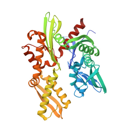
5AR0

HSP72 with adenosine-derived inhibitor


Experimental Data Snapshot

  • Method: X-RAY DIFFRACTION
  • Resolution: 1.90 Å
  • R-Value Free: 0.210 
  • R-Value Work: 0.172 
  • R-Value Observed: 0.174 

Starting Model: experimental
View more details

wwPDB Validation   3D Report Full Report


Ligand Structure Quality Assessment 


This is version 1.2 of the entry. See complete history


Literature

Exploiting Protein Conformational Change to Optimize Adenosine-Derived Inhibitors of Hsp70.

Cheeseman, M.D.Westwood, I.M.Barbeau, O.Rowlands, M.G.Dobson, S.Jones, A.M.Jeganathan, F.Burke, R.Kadi, N.Workman, P.Collins, I.Van Montfort, R.L.M.Jones, K.

(2016) J Med Chem 59: 4625

  • DOI: https://doi.org/10.1021/acs.jmedchem.5b02001
  • Primary Citation of Related Structures:  
    5AQZ, 5AR0

  • PubMed Abstract: 

    HSP70 is a molecular chaperone and a key component of the heat-shock response. Because of its proposed importance in oncology, this protein has become a popular target for drug discovery, efforts which have as yet brought little success. This study demonstrates that adenosine-derived HSP70 inhibitors potentially bind to the protein with a novel mechanism of action, the stabilization by desolvation of an intramolecular salt-bridge which induces a conformational change in the protein, leading to high affinity ligands. We also demonstrate that through the application of this mechanism, adenosine-derived HSP70 inhibitors can be optimized in a rational manner.


  • Organizational Affiliation

    Cancer Research UK Cancer Therapeutics Unit, The Institute of Cancer Research , London SW7 3RP, U.K.


Macromolecules
Find similar proteins by:  (by identity cutoff)  |  3D Structure
Entity ID: 1
MoleculeChains Sequence LengthOrganismDetailsImage
HEAT SHOCK 70 KDA PROTEIN 1A394Homo sapiensMutation(s): 0 
EC: 3.6.3.51
UniProt & NIH Common Fund Data Resources
Find proteins for P0DMV8 (Homo sapiens)
Explore P0DMV8 
Go to UniProtKB:  P0DMV8
PHAROS:  P0DMV8
GTEx:  ENSG00000204389 
Entity Groups  
Sequence Clusters30% Identity50% Identity70% Identity90% Identity95% Identity100% Identity
UniProt GroupP0DMV8
Sequence Annotations
Expand
  • Reference Sequence
Small Molecules
Ligands 4 Unique
IDChains Name / Formula / InChI Key2D Diagram3D Interactions
GB8
Query on GB8

Download Ideal Coordinates CCD File 
B [auth A](2R,3R,4S,5R)-2-(6-amino-8-((quinolin-7-ylmethyl)amino)-9H-purin-9-yl)-5-(hydroxymethyl)tetrahydrofuran-3,4-diol
C20 H21 N7 O4
ZPAVHMFARWZWPX-NVQRDWNXSA-N
GOL
Query on GOL

Download Ideal Coordinates CCD File 
C [auth A],
D [auth A],
E [auth A]
GLYCEROL
C3 H8 O3
PEDCQBHIVMGVHV-UHFFFAOYSA-N
DMS
Query on DMS

Download Ideal Coordinates CCD File 
F [auth A],
G [auth A]
DIMETHYL SULFOXIDE
C2 H6 O S
IAZDPXIOMUYVGZ-UHFFFAOYSA-N
CL
Query on CL

Download Ideal Coordinates CCD File 
H [auth A]CHLORIDE ION
Cl
VEXZGXHMUGYJMC-UHFFFAOYSA-M
Binding Affinity Annotations 
IDSourceBinding Affinity
GB8 BindingDB:  5AR0 Ki: 1290 (nM) from 1 assay(s)
Kd: min: 720, max: 724 (nM) from 2 assay(s)
IC50: 2900 (nM) from 1 assay(s)
Experimental Data & Validation

Experimental Data

  • Method: X-RAY DIFFRACTION
  • Resolution: 1.90 Å
  • R-Value Free: 0.210 
  • R-Value Work: 0.172 
  • R-Value Observed: 0.174 
  • Space Group: P 21 21 21
Unit Cell:
Length ( Å )Angle ( ˚ )
a = 47.88α = 90
b = 80.545β = 90
c = 92.979γ = 90
Software Package:
Software NamePurpose
BUSTERrefinement
XDSdata reduction
Aimlessdata scaling
PHASERphasing

Structure Validation

View Full Validation Report


Ligand Structure Quality Assessment 


Entry History 

Revision History  (Full details and data files)

  • Version 1.0: 2016-05-11
    Type: Initial release
  • Version 1.1: 2016-06-08
    Changes: Database references
  • Version 1.2: 2024-01-10
    Changes: Data collection, Database references, Derived calculations, Other, Refinement description