5AVZ | pdb_00005avz

Kinetics by X-ray crystallography: Tl+-substitution of bound K+ in the E2.MgF42-.2K+ crystal after 55 min


Experimental Data Snapshot

  • Method: X-RAY DIFFRACTION
  • Resolution: 3.20 Å
  • R-Value Free: 
    0.298 (Depositor), 0.283 (DCC) 
  • R-Value Work: 
    0.297 (Depositor), 0.287 (DCC) 
  • R-Value Observed: 
    0.297 (Depositor) 

Starting Model: experimental
View more details

wwPDB Validation 3D Report Full Report

Validation slider image for 5AVZ

Ligand Structure Quality Assessment 


This is version 2.2 of the entry. See complete history

Literature

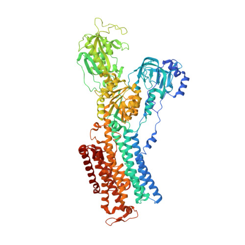
Sequential substitution of K(+) bound to Na(+),K(+)-ATPase visualized by X-ray crystallography.

Ogawa, H.Cornelius, F.Hirata, A.Toyoshima, C.

(2015) Nat Commun 6: 8004-8004

  • DOI: https://doi.org/10.1038/ncomms9004
  • Primary Citation Related Structures: 
    5AVQ, 5AVR, 5AVS, 5AVT, 5AVU, 5AVV, 5AVW, 5AVX, 5AVY, 5AVZ, 5AW0, 5AW1, 5AW2, 5AW3, 5AW4, 5AW5, 5AW6, 5AW7, 5AW8, 5AW9

  • PubMed Abstract: 

    Na(+),K(+)-ATPase transfers three Na(+) from the cytoplasm into the extracellular medium and two K(+) in the opposite direction per ATP hydrolysed. The binding and release of Na(+) and K(+) are all thought to occur sequentially. Here we demonstrate by X-ray crystallography of the ATPase in E2·MgF4(2-)·2K(+), a state analogous to E2·Pi·2K(+), combined with isotopic measurements, that the substitution of the two K(+) with congeners in the extracellular medium indeed occurs at different rates, substantially faster at site II. An analysis of thermal movements of protein atoms in the crystal shows that the M3-M4E helix pair opens and closes the ion pathway leading to the extracellular medium, allowing K(+) at site II to be substituted first. Taken together, these results indicate that site I K(+) is the first cation to bind to the empty cation-binding sites after releasing three Na(+).


  • Organizational Affiliation
    • Institute of Molecular and Cellular Biosciences, The University of Tokyo, Bunkyo-ku, Tokyo 113-0032, Japan.

Macromolecule Content 

  • Total Structure Weight: 158.52 kDa 
  • Atom Count: 10,235 
  • Modeled Residue Count: 1,299 
  • Deposited Residue Count: 1,407 
  • Unique protein chains: 3

Macromolecules

Find similar proteins by:|  3D Structure
Entity ID: 1
MoleculeChains  Sequence LengthOrganismDetailsImage
Na, K-ATPase alpha subunit1,028Squalus acanthiasMutation(s): 0 
Membrane Entity: Yes 
UniProt
Find proteins for Q4H132 (Squalus acanthias)
Explore Q4H132 
Go to UniProtKB:  Q4H132
Entity Groups
Sequence Clusters30% Identity50% Identity70% Identity90% Identity95% Identity100% Identity
UniProt GroupQ4H132
Sequence Annotations
Expand
Reference Sequence
Find similar proteins by:|  3D Structure
Entity ID: 2
MoleculeChains  Sequence LengthOrganismDetailsImage
Na+,K+-ATPase beta subunit305Squalus acanthiasMutation(s): 0 
Membrane Entity: Yes 
UniProt
Find proteins for C4IX13 (Squalus acanthias)
Explore C4IX13 
Go to UniProtKB:  C4IX13
Entity Groups
Sequence Clusters30% Identity50% Identity70% Identity90% Identity95% Identity100% Identity
UniProt GroupC4IX13
Glycosylation
Glycosylation Sites: 2
Sequence Annotations
Expand
Reference Sequence
Find similar proteins by:|  3D Structure
Entity ID: 3
MoleculeChains  Sequence LengthOrganismDetailsImage
Phospholemman-like proteinC [auth G]74Squalus acanthiasMutation(s): 0 
Membrane Entity: Yes 
UniProt
Find proteins for Q70Q12 (Squalus acanthias)
Explore Q70Q12 
Go to UniProtKB:  Q70Q12
Entity Groups
Sequence Clusters30% Identity50% Identity70% Identity90% Identity95% Identity100% Identity
UniProt GroupQ70Q12
Sequence Annotations
Expand
Reference Sequence

Oligosaccharides

Help  
Entity ID: 4
MoleculeChains Length2D Diagram GlycosylationD Interactions
2-acetamido-2-deoxy-beta-D-glucopyranose-(1-4)-2-acetamido-2-deoxy-beta-D-glucopyranoseD [auth C]2N-Glycosylation
Glycosylation Resources
GlyTouCan: G42666HT
GlyCosmos: G42666HT
GlyGen: G42666HT

Small Molecules

Ligands 6 Unique
IDChains Name / Formula / InChI Key2D Diagram3D Interactions
CLR

Query on CLR


Download:Ideal Coordinates CCD File
K [auth B]CHOLESTEROL
C27 H46 O
HVYWMOMLDIMFJA-DPAQBDIFSA-N
NAG

Query on NAG


Download:Ideal Coordinates CCD File
L [auth B]2-acetamido-2-deoxy-beta-D-glucopyranose
C8 H15 N O6
OVRNDRQMDRJTHS-FMDGEEDCSA-N
TL

Query on TL


Download:Ideal Coordinates CCD File
G [auth A],
H [auth A],
J [auth A]
THALLIUM (I) ION
Tl
ZLUSCZLCHQSJRU-UHFFFAOYSA-N
MF4

Query on MF4


Download:Ideal Coordinates CCD File
E [auth A]TETRAFLUOROMAGNESATE(2-)
F4 Mg
XVYWAXYEHHUKQW-UHFFFAOYSA-J
K

Query on K


Download:Ideal Coordinates CCD File
I [auth A]POTASSIUM ION
K
NPYPAHLBTDXSSS-UHFFFAOYSA-N
MG

Query on MG


Download:Ideal Coordinates CCD File
F [auth A]MAGNESIUM ION
Mg
JLVVSXFLKOJNIY-UHFFFAOYSA-N

Experimental Data & Validation

Experimental Data

  • Method: X-RAY DIFFRACTION
  • Resolution: 3.20 Å
  • R-Value Free:  0.298 (Depositor), 0.283 (DCC) 
  • R-Value Work:  0.297 (Depositor), 0.287 (DCC) 
  • R-Value Observed: 0.297 (Depositor) 
Space Group: C 1 2 1
Unit Cell:
Length ( Å )Angle ( ˚ )
a = 222.704α = 90
b = 50.914β = 104.52
c = 164.695γ = 90
Software Package:
Software NamePurpose
CNSrefinement
DENZOdata reduction
SCALEPACKdata scaling
CNSphasing

Structure Validation

View Full Validation Report


Ligand Structure Quality Assessment 


Entry History 

& Funding Information

Deposition Data


Funding OrganizationLocationGrant Number
Ministry of Education, Culture, Sports, Science and Technology of JapanJapan--
The Danish Medical Research FoundationDenmark--
The Danish Agency for Science Technology and InnovationDenmark--
The Novo Nordisk FoundationDenmark--

Revision History  (Full details and data files)

  • Version 1.0: 2015-09-02
    Type: Initial release
  • Version 1.1: 2020-02-26
    Changes: Data collection, Derived calculations, Source and taxonomy
  • Version 2.0: 2020-07-29
    Type: Remediation
    Reason: Carbohydrate remediation
    Changes: Atomic model, Data collection, Derived calculations, Structure summary
  • Version 2.1: 2023-11-08
    Changes: Data collection, Database references, Refinement description, Structure summary
  • Version 2.2: 2024-11-06
    Changes: Structure summary