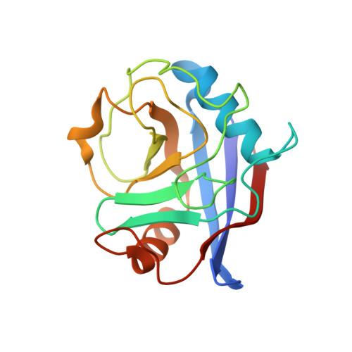
5CYH

CYCLOPHILIN A COMPLEXED WITH DIPEPTIDE GLY-PRO


Experimental Data Snapshot

  • Method: X-RAY DIFFRACTION
  • Resolution: 2.10 Å
  • R-Value Work: 0.190 
  • R-Value Observed: 0.190 

wwPDB Validation   3D Report Full Report


This is version 1.4 of the entry. See complete history


Literature

Mechanistic implication of crystal structures of the cyclophilin-dipeptide complexes.

Zhao, Y.Ke, H.

(1996) Biochemistry 35: 7362-7368

  • DOI: https://doi.org/10.1021/bi960278x
  • Primary Citation of Related Structures:  
    2CYH, 3CYH, 4CYH, 5CYH

  • PubMed Abstract: 

    The structures of cyclophilin A complexed with dipeptides of Ser-Pro, His-Pro, and Gly-Pro have been determined and refined at high resolution. Comparison of these structures revealed that the dipeptide complexes have the same molecular conformation and the same binding of the dipeptides. The side chains of the N-terminal amino acid of the above dipeptides do not strongly interact with cyclophilin, implying their minor contribution to the cis-trans isomerization and thus accounting for the broad catalytic specificity of the enzyme. The binding of the dipeptides is similar to that of the common substrate succinyl-Ala-Ala-Pro-Phe-p-nitroanilide in terms of the N-terminal hydrogen bonding and the hydrophobic interaction of the proline side chain. However, substantial difference between these structures are observed in (1) hydrogen bonding between the carboxyl terminus of the peptides and Arg55 and between Arg55 and Gln63, (2) the side chain conformation of Arg55, and (3) water binding at the active site. These differences imply either that dipeptides are not substrates but competitive inhibitors of peptidyl-prolyl cis-trans isomerases or that dipeptides are subject to different catalytic mechanisms from tetrapeptides.


  • Organizational Affiliation

    Department of Biochemistry and Biophysics School of Medicine, University of North Carolina, Chapel Hill 27599, USA.


Macromolecules
Find similar proteins by:  (by identity cutoff)  |  3D Structure
Entity ID: 1
MoleculeChains Sequence LengthOrganismDetailsImage
CYCLOPHILIN A164Homo sapiensMutation(s): 0 
Gene Names: CYCLOPHILIN
EC: 5.2.1.8
UniProt & NIH Common Fund Data Resources
Find proteins for P62937 (Homo sapiens)
Explore P62937 
Go to UniProtKB:  P62937
PHAROS:  P62937
GTEx:  ENSG00000196262 
Entity Groups  
Sequence Clusters30% Identity50% Identity70% Identity90% Identity95% Identity100% Identity
UniProt GroupP62937
Sequence Annotations
Expand
  • Reference Sequence
Small Molecules
Ligands 2 Unique
IDChains Name / Formula / InChI Key2D Diagram3D Interactions
PRO
Query on PRO

Download Ideal Coordinates CCD File 
C [auth A]PROLINE
C5 H9 N O2
ONIBWKKTOPOVIA-BYPYZUCNSA-N
GLY
Query on GLY

Download Ideal Coordinates CCD File 
B [auth A]GLYCINE
C2 H5 N O2
DHMQDGOQFOQNFH-UHFFFAOYSA-N
Experimental Data & Validation

Experimental Data

  • Method: X-RAY DIFFRACTION
  • Resolution: 2.10 Å
  • R-Value Work: 0.190 
  • R-Value Observed: 0.190 
  • Space Group: P 21 21 21
Unit Cell:
Length ( Å )Angle ( ˚ )
a = 40.8α = 90
b = 52.4β = 90
c = 90.1γ = 90
Software Package:
Software NamePurpose
X-PLORrefinement
RIGAKUdata reduction
RIGAKUdata scaling

Structure Validation

View Full Validation Report


Entry History 

Deposition Data

Revision History  (Full details and data files)

  • Version 1.0: 1996-07-11
    Type: Initial release
  • Version 1.1: 2008-03-25
    Changes: Version format compliance
  • Version 1.2: 2011-07-13
    Changes: Version format compliance
  • Version 1.3: 2017-11-29
    Changes: Derived calculations, Other
  • Version 1.4: 2024-03-06
    Changes: Data collection, Database references, Derived calculations