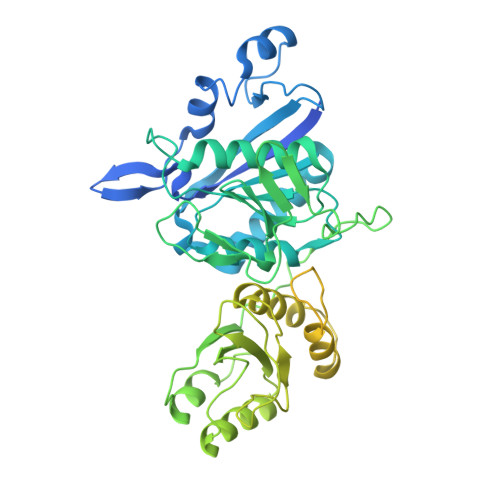
5DNC

Crystal structure of the Asn-bound guinea pig L-asparaginase 1 catalytic domain active site mutant T19A

  • Classification: HYDROLASE
  • Organism(s): Cavia porcellus
  • Expression System: Escherichia coli
  • Mutation(s): Yes 

  • Deposited: 2015-09-09 Released: 2016-01-13 
  • Deposition Author(s): Schalk, A.M., Lavie, A.
  • Funding Organization(s): National Institutes of Health/National Institute of Biomedical Imaging and Bioengineering (NIH/NIBIB), Department of Veterans Affairs Biomedical Laboratory Research and Development Service

Experimental Data Snapshot

  • Method: X-RAY DIFFRACTION
  • Resolution: 2.01 Å
  • R-Value Free: 0.212 
  • R-Value Work: 0.187 
  • R-Value Observed: 0.188 

Starting Model: experimental
View more details

wwPDB Validation   3D Report Full Report


This is version 1.6 of the entry. See complete history


Literature

Experimental Data in Support of a Direct Displacement Mechanism for Type I/II l-Asparaginases.

Schalk, A.M.Antansijevic, A.Caffrey, M.Lavie, A.

(2016) J Biol Chem 291: 5088-5100

  • DOI: https://doi.org/10.1074/jbc.M115.699884
  • Primary Citation of Related Structures:  
    5DNC, 5DND, 5DNE

  • PubMed Abstract: 

    Bacterial L-asparaginases play an important role in the treatment of certain types of blood cancers. We are exploring the guinea pig L-asparaginase (gpASNase1) as a potential replacement of the immunogenic bacterial enzymes. The exact mechanism used by L-asparaginases to catalyze the hydrolysis of asparagine into aspartic acid and ammonia has been recently put into question. Earlier experimental data suggested that the reaction proceeds via a covalent intermediate using a ping-pong mechanism, whereas recent computational work advocates the direct displacement of the amine by an activated water. To shed light on this controversy, we generated gpASNase1 mutants of conserved active site residues (T19A, T116A, T19A/T116A, K188M, and Y308F) suspected to play a role in hydrolysis. Using x-ray crystallography, we determined the crystal structures of the T19A, T116A, and K188M mutants soaked in asparagine. We also characterized their steady-state kinetic properties and analyzed the conversion of asparagine to aspartate using NMR. Our structures reveal bound asparagine in the active site that has unambiguously not formed a covalent intermediate. Kinetic and NMR assays detect significant residual activity for all of the mutants. Furthermore, no burst of ammonia production was observed that would indicate covalent intermediate formation and the presence of a ping-pong mechanism. Hence, despite using a variety of techniques, we were unable to obtain experimental evidence that would support the formation of a covalent intermediate. Consequently, our observations support a direct displacement rather than a ping-pong mechanism for l-asparaginases.


  • Organizational Affiliation

    From the Department of Biochemistry and Molecular Genetics, University of Illinois at Chicago, Chicago, Illinois 60607 and.


Macromolecules
Find similar proteins by:  (by identity cutoff)  |  3D Structure
Entity ID: 1
MoleculeChains Sequence LengthOrganismDetailsImage
L-asparaginase
A, B, C, D
588Cavia porcellusMutation(s): 1 
Gene Names: ASPG
EC: 3.5.1.1
UniProt
Find proteins for H0W0T5 (Cavia porcellus)
Explore H0W0T5 
Go to UniProtKB:  H0W0T5
Entity Groups  
Sequence Clusters30% Identity50% Identity70% Identity90% Identity95% Identity100% Identity
UniProt GroupH0W0T5
Sequence Annotations
Expand
  • Reference Sequence
Small Molecules
Ligands 2 Unique
IDChains Name / Formula / InChI Key2D Diagram3D Interactions
ASN
Query on ASN

Download Ideal Coordinates CCD File 
E [auth A],
K [auth B],
P [auth C],
U [auth D]
ASPARAGINE
C4 H8 N2 O3
DCXYFEDJOCDNAF-REOHCLBHSA-N
EDO
Query on EDO

Download Ideal Coordinates CCD File 
F [auth A]
G [auth A]
H [auth A]
I [auth A]
J [auth A]
F [auth A],
G [auth A],
H [auth A],
I [auth A],
J [auth A],
L [auth B],
M [auth B],
N [auth B],
O [auth B],
Q [auth C],
R [auth C],
S [auth C],
T [auth C],
V [auth D],
W [auth D],
X [auth D],
Y [auth D]
1,2-ETHANEDIOL
C2 H6 O2
LYCAIKOWRPUZTN-UHFFFAOYSA-N
Experimental Data & Validation

Experimental Data

  • Method: X-RAY DIFFRACTION
  • Resolution: 2.01 Å
  • R-Value Free: 0.212 
  • R-Value Work: 0.187 
  • R-Value Observed: 0.188 
  • Space Group: I 2 2 2
Unit Cell:
Length ( Å )Angle ( ˚ )
a = 123.19α = 90
b = 155β = 90
c = 157.1γ = 90
Software Package:
Software NamePurpose
REFMACrefinement
XDSdata reduction
XSCALEdata scaling
Cootmodel building

Structure Validation

View Full Validation Report


Entry History & Funding Information

Deposition Data


Funding OrganizationLocationGrant Number
National Institutes of Health/National Institute of Biomedical Imaging and Bioengineering (NIH/NIBIB)United StatesRO1 EB013685
Department of Veterans Affairs Biomedical Laboratory Research and Development ServiceUnited StatesMerit Review Award # I01BX001919

Revision History  (Full details and data files)

  • Version 1.0: 2016-01-13
    Type: Initial release
  • Version 1.1: 2016-01-20
    Changes: Database references
  • Version 1.2: 2016-03-23
    Changes: Database references
  • Version 1.3: 2017-09-27
    Changes: Author supporting evidence, Database references, Derived calculations
  • Version 1.4: 2017-11-01
    Changes: Author supporting evidence
  • Version 1.5: 2019-12-11
    Changes: Author supporting evidence
  • Version 1.6: 2023-09-27
    Changes: Data collection, Database references, Refinement description