5DUQ | pdb_00005duq

Active human c1-inhibitor in complex with dextran sulfate


Experimental Data Snapshot

  • Method: X-RAY DIFFRACTION
  • Resolution: 2.90 Å
  • R-Value Free: 
    0.278 (Depositor), 0.287 (DCC) 
  • R-Value Work: 
    0.228 (Depositor), 0.241 (DCC) 
  • R-Value Observed: 
    0.231 (Depositor) 

wwPDB Validation 3D Report Full Report

Validation slider image for 5DUQ

Ligand Structure Quality Assessment 


This is version 2.1 of the entry. See complete history

Literature

How Dextran Sulfate Affects C1-inhibitor Activity: A Model for Polysaccharide Potentiation.

Dijk, M.Holkers, J.Voskamp, P.Giannetti, B.M.Waterreus, W.J.van Veen, H.A.Pannu, N.S.

(2016) Structure 24: 2182-2189

  • DOI: https://doi.org/10.1016/j.str.2016.09.013
  • Primary Citation Related Structures: 
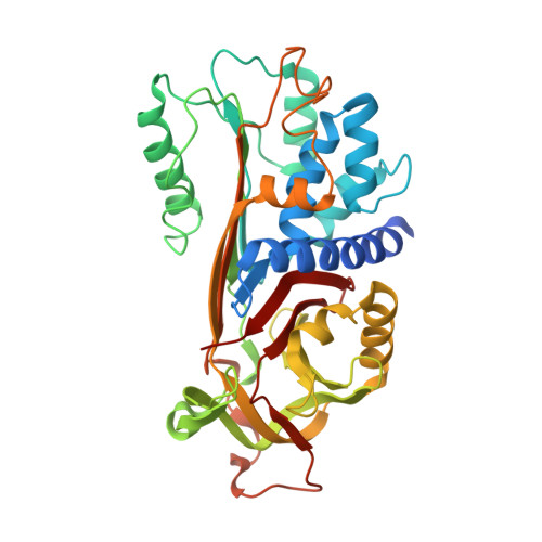
    5DU3, 5DUQ

  • PubMed Abstract: 

    C1-inhibitor is a key inhibitor of the complement and contact activation systems, and mutations in the protein can cause hereditary angioedema. Through an unknown mechanism, polysaccharides can increase C1-inhibitor activity against some of its target proteases. Here we present the crystal structures of the serine protease inhibitor (serpin) domain of active C1-inhibitor by itself and in complex with dextran sulfate. Unlike previously described interactions of serpins with polysaccharides, the structures and isothermal titration calorimetry experiments together reveal that dextran sulfate binds to C1-inhibitor's F1 helix with low affinity and does not invoke an allosteric change. Furthermore, one dextran sulfate molecule can bind multiple C1-inhibitor molecules. We propose that in a C1-inhibitor/protease/polysaccharide ternary complex, negatively charged polysaccharides link C1-inhibitor's positively charged F1 helix to positively charged autolysis loops of proteases. The proposed mechanism elegantly explains previous experiments showing that polysaccharide potentiation is increased against proteases with a greater positive charge in their autolysis loop.


  • Organizational Affiliation
    • Department of Biophysical Structural Chemistry, Gorlaeus Laboratories, Leiden University, Einsteinweg 55, 2333 CC Leiden, the Netherlands.

Macromolecule Content 

  • Total Structure Weight: 86.87 kDa 
  • Atom Count: 5,840 
  • Modeled Residue Count: 727 
  • Deposited Residue Count: 766 
  • Unique protein chains: 1

Macromolecules

Find similar proteins by:|  3D Structure
Entity ID: 1
MoleculeChains  Sequence LengthOrganismDetailsImage
Plasma protease C1 inhibitor
A, B
383Homo sapiensMutation(s): 0 
Gene Names: SERPING1C1INC1NH
UniProt & NIH Common Fund Data Resources
Find proteins for P05155 (Homo sapiens)
Explore P05155 
Go to UniProtKB:  P05155
PHAROS:  P05155
GTEx:  ENSG00000149131 
Entity Groups
Sequence Clusters30% Identity50% Identity70% Identity90% Identity95% Identity100% Identity
UniProt GroupP05155
Glycosylation
Glycosylation Sites: 1Go to GlyGen: P05155-1
Sequence Annotations
Expand
Reference Sequence

Oligosaccharides

Help  
Entity ID: 2
MoleculeChains Length2D Diagram GlycosylationD Interactions
beta-D-glucopyranose-(1-4)-alpha-D-glucopyranose
C
2N/A
Glycosylation Resources
GlyTouCan: G86802XL
GlyCosmos: G86802XL

Biologically Interesting Molecules (External Reference) 

1 Unique

Experimental Data & Validation

Experimental Data

  • Method: X-RAY DIFFRACTION
  • Resolution: 2.90 Å
  • R-Value Free:  0.278 (Depositor), 0.287 (DCC) 
  • R-Value Work:  0.228 (Depositor), 0.241 (DCC) 
  • R-Value Observed: 0.231 (Depositor) 
Space Group: C 1 2 1
Unit Cell:
Length ( Å )Angle ( ˚ )
a = 112.1α = 90
b = 197.424β = 103.31
c = 56.779γ = 90
Software Package:
Software NamePurpose
REFMACrefinement
XDSdata reduction
Aimlessdata scaling
MOLREPphasing

Structure Validation

View Full Validation Report


Ligand Structure Quality Assessment 


Entry History 

Deposition Data

Revision History  (Full details and data files)

  • Version 1.0: 2016-08-31
    Type: Initial release
  • Version 1.1: 2017-01-25
    Changes: Database references
  • Version 2.0: 2020-07-29
    Type: Remediation
    Reason: Carbohydrate remediation
    Changes: Atomic model, Data collection, Derived calculations, Structure summary
  • Version 2.1: 2024-10-23
    Changes: Data collection, Database references, Derived calculations, Refinement description, Structure summary