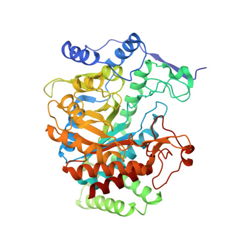
5E2L

3-deoxy-D-arabino-heptulosonate 7-phosphate synthase from Mycobacterium tuberculosis in complex with D-phenylalanine


Experimental Data Snapshot

  • Method: X-RAY DIFFRACTION
  • Resolution: 2.50 Å
  • R-Value Free: 0.174 
  • R-Value Work: 0.156 
  • R-Value Observed: 0.157 

Starting Model: experimental
View more details

wwPDB Validation   3D Report Full Report


Ligand Structure Quality Assessment 


This is version 1.1 of the entry. See complete history


Literature

Probing the Sophisticated Synergistic Allosteric Regulation of Aromatic Amino Acid Biosynthesis in Mycobacterium tuberculosis Using -Amino Acids.

Reichau, S.Blackmore, N.J.Jiao, W.Parker, E.J.

(2016) PLoS One 11: e0152723-e0152723

  • DOI: https://doi.org/10.1371/journal.pone.0152723
  • Primary Citation of Related Structures:  
    5E2L, 5E40, 5E4N, 5E5G, 5E7Z

  • PubMed Abstract: 

    Chirality plays a major role in recognition and interaction of biologically important molecules. The enzyme 3-deoxy-d-arabino-heptulosonate 7-phosphate synthase (DAH7PS) is the first enzyme of the shikimate pathway, which is responsible for the synthesis of aromatic amino acids in bacteria and plants, and a potential target for the development of antibiotics and herbicides. DAH7PS from Mycobacterium tuberculosis (MtuDAH7PS) displays an unprecedented complexity of allosteric regulation, with three interdependent allosteric binding sites and a ternary allosteric response to combinations of the aromatic amino acids l-Trp, l-Phe and l-Tyr. In order to further investigate the intricacies of this system and identify key residues in the allosteric network of MtuDAH7PS, we studied the interaction of MtuDAH7PS with aromatic amino acids that bear the non-natural d-configuration, and showed that the d-amino acids do not elicit an allosteric response. We investigated the binding mode of d-amino acids using X-ray crystallography, site directed mutagenesis and isothermal titration calorimetry. Key differences in the binding mode were identified: in the Phe site, a hydrogen bond between the amino group of the allosteric ligands to the side chain of Asn175 is not established due to the inverted configuration of the ligands. In the Trp site, d-Trp forms no interaction with the main chain carbonyl group of Thr240 and less favourable interactions with Asn237 when compared to the l-Trp binding mode. Investigation of the MtuDAH7PSN175A variant further supports the hypothesis that the lack of key interactions in the binding mode of the aromatic d-amino acids are responsible for the absence of an allosteric response, which gives further insight into which residues of MtuDAH7PS play a key role in the transduction of the allosteric signal.


  • Organizational Affiliation

    Biomolecular Interaction Centre and Department of Chemistry, University of Canterbury, Christchurch, New Zealand.


Macromolecules
Find similar proteins by:  (by identity cutoff)  |  3D Structure
Entity ID: 1
MoleculeChains Sequence LengthOrganismDetailsImage
3-deoxy-D-arabinoheptulosonate-7-phosphate synthase
A, B
464Mycobacterium tuberculosisMutation(s): 0 
Gene Names: aroG_1ERS024751_03564ERS094182_00944ERS124362_02783
EC: 2.5.1.54
UniProt
Find proteins for O53512 (Mycobacterium tuberculosis (strain ATCC 25618 / H37Rv))
Explore O53512 
Go to UniProtKB:  O53512
Entity Groups  
Sequence Clusters30% Identity50% Identity70% Identity90% Identity95% Identity100% Identity
UniProt GroupO53512
Sequence Annotations
Expand
  • Reference Sequence
Small Molecules
Ligands 5 Unique
IDChains Name / Formula / InChI Key2D Diagram3D Interactions
DPN
Query on DPN

Download Ideal Coordinates CCD File 
I [auth A],
R [auth B]
D-PHENYLALANINE
C9 H11 N O2
COLNVLDHVKWLRT-MRVPVSSYSA-N
SO4
Query on SO4

Download Ideal Coordinates CCD File 
C [auth A],
J [auth B],
K [auth B],
L [auth B],
M [auth B]
SULFATE ION
O4 S
QAOWNCQODCNURD-UHFFFAOYSA-L
GOL
Query on GOL

Download Ideal Coordinates CCD File 
F [auth A],
P [auth B]
GLYCEROL
C3 H8 O3
PEDCQBHIVMGVHV-UHFFFAOYSA-N
MN
Query on MN

Download Ideal Coordinates CCD File 
D [auth A],
N [auth B]
MANGANESE (II) ION
Mn
WAEMQWOKJMHJLA-UHFFFAOYSA-N
CL
Query on CL

Download Ideal Coordinates CCD File 
E [auth A],
G [auth A],
H [auth A],
O [auth B],
Q [auth B]
CHLORIDE ION
Cl
VEXZGXHMUGYJMC-UHFFFAOYSA-M
Experimental Data & Validation

Experimental Data

  • Method: X-RAY DIFFRACTION
  • Resolution: 2.50 Å
  • R-Value Free: 0.174 
  • R-Value Work: 0.156 
  • R-Value Observed: 0.157 
  • Space Group: P 32 2 1
Unit Cell:
Length ( Å )Angle ( ˚ )
a = 203.989α = 90
b = 203.989β = 90
c = 66.904γ = 120
Software Package:
Software NamePurpose
REFMACrefinement
Aimlessdata scaling
PDB_EXTRACTdata extraction
XDSdata reduction
MOLREPphasing

Structure Validation

View Full Validation Report


Ligand Structure Quality Assessment 


Entry History & Funding Information

Deposition Data


Funding OrganizationLocationGrant Number
New Zealand Marsden FundNew ZealandUOC1105
Maurice Wilkins Centre for Molecular BiodiscoveryNew Zealand--

Revision History  (Full details and data files)

  • Version 1.0: 2016-06-01
    Type: Initial release
  • Version 1.1: 2023-09-27
    Changes: Data collection, Database references, Derived calculations, Refinement description