5FKJ

Crystal structure of mouse acetylcholinesterase in complex with C-547, an alkyl ammonium derivative of 6-methyl uracil


Experimental Data Snapshot

  • Method: X-RAY DIFFRACTION
  • Resolution: 3.13 Å
  • R-Value Free: 
    0.249 (Depositor), 0.250 (DCC) 
  • R-Value Work: 
    0.207 (Depositor), 0.210 (DCC) 
  • R-Value Observed: 
    0.208 (Depositor) 

Starting Model: experimental
View more details

wwPDB Validation   3D Report Full Report


Ligand Structure Quality Assessment 

Created with Raphaël 2.3.0Worse 01 BetterLigand structure goodness of fit to experimental dataBest fitted G0WClick on this verticalbar to view detailsBest fitted NAGClick on this verticalbar to view details

This is version 1.5 of the entry. See complete history


Literature

Slow-Binding Inhibition of Acetylcholinesterase by a 6-Methyluracil Alkyl-Ammonium Derivative: Mechanism and Advantages for Myasthenia Gravis Treatment.

Kharlamova, A.D.Lushchekina, S.V.Petrov, K.A.Kots, E.D.Nachon, F.V.Villard-Wandhammer, M.Zueva, I.V.Krejci, E.Reznik, V.S.Zobov, V.V.Nikolsky, E.E.Masson, P.

(2016) Biochem J 473: 1225

  • DOI: https://doi.org/10.1042/BCJ20160084
  • Primary Citation of Related Structures:  
    5FKJ

  • PubMed Abstract: 

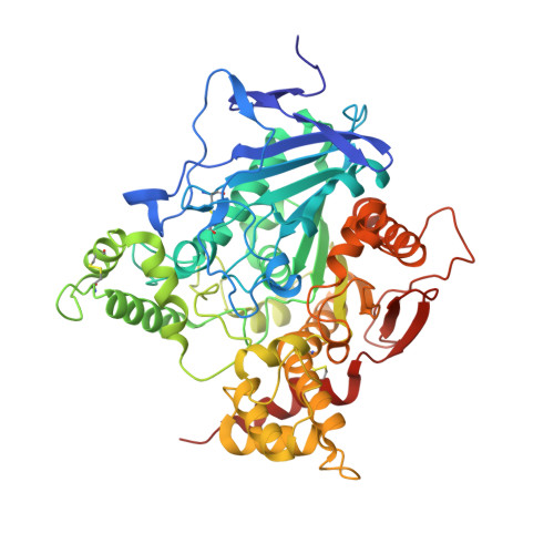
    Inhibition of human AChE (acetylcholinesterase) and BChE (butyrylcholinesterase) by an alkylammonium derivative of 6-methyluracil, C-547, a potential drug for the treatment of MG (myasthenia gravis) was studied. Kinetic analysis of AChE inhibition showed that C-547 is a slow-binding inhibitor of type B, i.e. after formation of the initial enzyme·inhibitor complex (Ki=140 pM), an induced-fit step allows establishment of the final complex (Ki*=22 pM). The estimated koff is low, 0.05 min(-1) On the other hand, reversible inhibition of human BChE is a fast-binding process of mixed-type (Ki=1.77 μM; Ki'=3.17 μM). The crystal structure of mouse AChE complexed with C-547 was solved at 3.13 Å resolution. The complex is stabilized by cation-π, stacking and hydrogen-bonding interactions. Molecular dynamics simulations of the binding/dissociation processes of C-547 and C-35 (a non-charged analogue) to mouse and human AChEs were performed. Molecular modelling on mouse and human AChE showed that the slow step results from an enzyme conformational change that allows C-547 to cross the bottleneck in the active-site gorge, followed by formation of tight complex, as observed in the crystal structure. In contrast, the related non-charged compound C-35 is not a slow-binding inhibitor. It does not cross the bottleneck because it is not sensitive to the electrostatic driving force to reach the bottom of the gorge. Thus C-547 is one of the most potent and selective reversible inhibitors of AChE with a long residence time, τ=20 min, longer than for other reversible inhibitors used in the treatment of MG. This makes C-547 a promising drug for the treatment of this disease.


  • Organizational Affiliation

    A.E. Arbuzov Institute of Organic and Physical Chemistry of Russian Academy of Sciences, Arbuzov Str. 8, Kazan 420088 Russia.


Macromolecules
Find similar proteins by:  (by identity cutoff)  |  3D Structure
Entity ID: 1
MoleculeChains Sequence LengthOrganismDetailsImage
ACETYLCHOLINESTERASE
A, B, C, D
543Mus musculusMutation(s): 0 
EC: 3.1.1.7
UniProt & NIH Common Fund Data Resources
Find proteins for P21836 (Mus musculus)
Explore P21836 
Go to UniProtKB:  P21836
IMPC:  MGI:87876
Entity Groups  
Sequence Clusters30% Identity50% Identity70% Identity90% Identity95% Identity100% Identity
UniProt GroupP21836
Glycosylation
Glycosylation Sites: 1Go to GlyGen: P21836-1
Sequence Annotations
Expand
  • Reference Sequence
Small Molecules
Ligands 4 Unique
IDChains Name / Formula / InChI Key2D Diagram3D Interactions
G0W
Query on G0W

Download Ideal Coordinates CCD File 
E [auth A],
JA [auth D],
P [auth B],
X [auth C]
1,3-BIS[5(DIETHYL-O-NITROBENZYLAMMONIUM)PENTYL]-6-METHYLURACIL
C37 H56 N6 O6
WLTAFWKKNBLBHO-UHFFFAOYSA-N
NAG
Query on NAG

Download Ideal Coordinates CCD File 
F [auth A]
KA [auth D]
LA [auth D]
Q [auth B]
R [auth B]
2-acetamido-2-deoxy-beta-D-glucopyranose
C8 H15 N O6
OVRNDRQMDRJTHS-FMDGEEDCSA-N
SO4
Query on SO4

Download Ideal Coordinates CCD File 
IA [auth C],
SA [auth D],
W [auth B]
SULFATE ION
O4 S
QAOWNCQODCNURD-UHFFFAOYSA-L
CL
Query on CL

Download Ideal Coordinates CCD File 
AA [auth C]
BA [auth C]
CA [auth C]
DA [auth C]
EA [auth C]
CHLORIDE ION
Cl
VEXZGXHMUGYJMC-UHFFFAOYSA-M
Experimental Data & Validation

Experimental Data

  • Method: X-RAY DIFFRACTION
  • Resolution: 3.13 Å
  • R-Value Free:  0.249 (Depositor), 0.250 (DCC) 
  • R-Value Work:  0.207 (Depositor), 0.210 (DCC) 
  • R-Value Observed: 0.208 (Depositor) 
Space Group: P 21 21 21
Unit Cell:
Length ( Å )Angle ( ˚ )
a = 137.15α = 90
b = 175.45β = 90
c = 224.4γ = 90
Software Package:
Software NamePurpose
PHENIXrefinement
XDSdata reduction
XSCALEdata scaling
MOLREPphasing

Structure Validation

View Full Validation Report


Ligand Structure Quality Assessment 

Created with Raphaël 2.3.0Worse 01 BetterLigand structure goodness of fit to experimental dataBest fitted G0WClick on this verticalbar to view detailsBest fitted NAGClick on this verticalbar to view details

Entry History 

Deposition Data

Revision History  (Full details and data files)

  • Version 1.0: 2016-03-16
    Type: Initial release
  • Version 1.1: 2016-05-11
    Changes: Database references
  • Version 1.2: 2017-08-09
    Changes: Data collection
  • Version 1.3: 2020-07-29
    Type: Remediation
    Reason: Carbohydrate remediation
    Changes: Data collection, Derived calculations, Other, Structure summary
  • Version 1.4: 2024-01-10
    Changes: Data collection, Database references, Refinement description, Structure summary
  • Version 1.5: 2024-11-13
    Changes: Structure summary