5FVM | pdb_00005fvm

Cryo electron microscopy of a complex of Tor and Lst8


Experimental Data Snapshot

  • Method: ELECTRON MICROSCOPY
  • Resolution: 6.70 Å
  • Aggregation State: PARTICLE 
  • Reconstruction Method: SINGLE PARTICLE 

Starting Model: experimental
View more details

wwPDB Validation 3D Report Full Report

Validation slider image for 5FVM

This is version 1.4 of the entry. See complete history

Literature

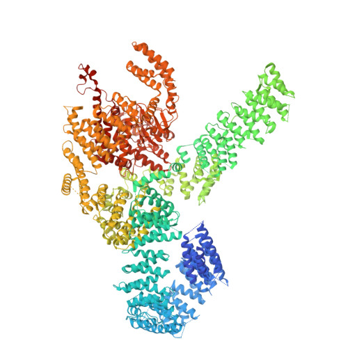
Tor Forms a Dimer Through an N-Terminal Helical Solenoid with a Complex Topology

Baretic, D.Berndt, A.Ohashi, Y.Johnson, C.M.Williams, R.L.

(2016) Nat Commun 7: 11016

  • DOI: https://doi.org/10.1038/ncomms11016
  • Primary Citation Related Structures: 
    5FVM

  • PubMed Abstract: 

    The target of rapamycin (Tor) is a Ser/Thr protein kinase that regulates a range of anabolic and catabolic processes. Tor is present in two complexes, TORC1 and TORC2, in which the Tor-Lst8 heterodimer forms a common sub-complex. We have determined the cryo-electron microscopy (EM) structure of Tor bound to Lst8. Two Tor-Lst8 heterodimers assemble further into a dyad-symmetry dimer mediated by Tor-Tor interactions. The first 1,300 residues of Tor form a HEAT repeat-containing α-solenoid with four distinct segments: a highly curved 800-residue N-terminal 'spiral', followed by a 400-residue low-curvature 'bridge' and an extended 'railing' running along the bridge leading to the 'cap' that links to FAT region. This complex topology was verified by domain insertions and offers a new interpretation of the mTORC1 structure. The spiral of one TOR interacts with the bridge of another, which together form a joint platform for the Regulatory Associated Protein of TOR (RAPTOR) regulatory subunit.


  • Organizational Affiliation
    • MRC Laboratory of Molecular Biology, Cambridge CB2 0QH, UK.

Macromolecule Content 

  • Total Structure Weight: 626.72 kDa 
  • Atom Count: 23,974 
  • Modeled Residue Count: 4,832 
  • Deposited Residue Count: 5,548 
  • Unique protein chains: 2

Macromolecules

Find similar proteins by:|  3D Structure
Entity ID: 1
MoleculeChains  Sequence LengthOrganismDetailsImage
SERINE/THREONINE-PROTEIN KINASE TOR2
A, B
2,471Kluyveromyces marxianusMutation(s): 0 
Gene Names: KMAR_30040
Find similar proteins by:|  3D Structure
Entity ID: 2
MoleculeChains  Sequence LengthOrganismDetailsImage
TARGET OF RAPAMYCIN COMPLEX SUBUNIT LST8
C, D
303Kluyveromyces marxianusMutation(s): 0 
Gene Names: KMAR_20458

Experimental Data & Validation

Experimental Data

  • Method: ELECTRON MICROSCOPY
  • Resolution: 6.70 Å
  • Aggregation State: PARTICLE 
  • Reconstruction Method: SINGLE PARTICLE 
EM Software:
TaskSoftware PackageVersion
RECONSTRUCTIONRELION

Structure Validation

View Full Validation Report


Entry History 

Deposition Data

Revision History  (Full details and data files)

  • Version 1.0: 2016-04-20
    Type: Initial release
  • Version 1.1: 2016-04-27
    Changes: Database references
  • Version 1.2: 2017-08-02
    Changes: Data collection, Refinement description
  • Version 1.3: 2019-10-23
    Changes: Data collection, Other
  • Version 1.4: 2024-05-08
    Changes: Data collection, Database references, Refinement description