5HQ8

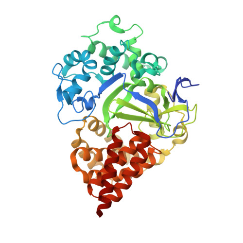
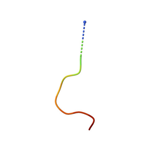
Co-crystal Structure of human SMYD3 with a MEKK2 peptide at 2.13A


Experimental Data Snapshot

  • Method: X-RAY DIFFRACTION
  • Resolution: 1.72 Å
  • R-Value Free: 0.201 
  • R-Value Work: 0.174 
  • R-Value Observed: 0.175 

wwPDB Validation   3D Report Full Report


Ligand Structure Quality Assessment 


This is version 1.4 of the entry. See complete history


Literature

Structure-Based Design of a Novel SMYD3 Inhibitor that Bridges the SAM-and MEKK2-Binding Pockets.

Van Aller, G.S.Graves, A.P.Elkins, P.A.Bonnette, W.G.McDevitt, P.J.Zappacosta, F.Annan, R.S.Dean, T.W.Su, D.S.Carpenter, C.L.Mohammad, H.P.Kruger, R.G.

(2016) Structure 24: 774-781

  • DOI: https://doi.org/10.1016/j.str.2016.03.010
  • Primary Citation of Related Structures:  
    5HI7, 5HQ8

  • PubMed Abstract: 

    SMYD3 is a lysine methyltransferase overexpressed in colorectal, breast, prostate, and hepatocellular tumors, and has been implicated as an oncogene in human malignancies. Methylation of MEKK2 by SMYD3 is important for regulation of the MEK/ERK pathway, suggesting the possibility of selectively targeting SMYD3 in RAS-driven cancers. Structural and kinetic characterization of SMYD3 was undertaken leading to a co-crystal structure of SMYD3 with a MEKK2-peptide substrate bound, and the observation that SMYD3 follows a partially processive mechanism. These insights allowed for the design of GSK2807, a potent and selective, SAM-competitive inhibitor of SMYD3 (Ki = 14 nM). A high-resolution crystal structure reveals that GSK2807 bridges the gap between the SAM-binding pocket and the substrate lysine tunnel of SMYD3. Taken together, our data demonstrate that small-molecule inhibitors of SMYD3 can be designed to prevent methylation of MEKK2 and these could have potential use as anticancer therapeutics.


  • Organizational Affiliation

    Cancer Epigenetics Discovery Performance Unit, GlaxoSmithKline, Collegeville, PA 19426, USA.


Macromolecules
Find similar proteins by:  (by identity cutoff)  |  3D Structure
Entity ID: 1
MoleculeChains Sequence LengthOrganismDetailsImage
Histone-lysine N-methyltransferase SMYD3
A, B
432Homo sapiensMutation(s): 0 
Gene Names: SMYD3ZMYND1ZNFN3A1
EC: 2.1.1.43 (PDB Primary Data), 2.1.1.354 (UniProt)
UniProt & NIH Common Fund Data Resources
Find proteins for Q9H7B4 (Homo sapiens)
Explore Q9H7B4 
Go to UniProtKB:  Q9H7B4
PHAROS:  Q9H7B4
GTEx:  ENSG00000185420 
Entity Groups  
Sequence Clusters30% Identity50% Identity70% Identity90% Identity95% Identity100% Identity
UniProt GroupQ9H7B4
Sequence Annotations
Expand
  • Reference Sequence

Find similar proteins by:  Sequence   |   3D Structure  

Entity ID: 2
MoleculeChains Sequence LengthOrganismDetailsImage
MEKK2 peptideC [auth I],
D [auth J]
16Homo sapiensMutation(s): 0 
EC: 2.7.11.25
UniProt & NIH Common Fund Data Resources
Find proteins for Q9Y2U5 (Homo sapiens)
Explore Q9Y2U5 
Go to UniProtKB:  Q9Y2U5
PHAROS:  Q9Y2U5
GTEx:  ENSG00000169967 
Entity Groups  
Sequence Clusters30% Identity50% Identity70% Identity90% Identity95% Identity100% Identity
UniProt GroupQ9Y2U5
Sequence Annotations
Expand
  • Reference Sequence
Small Molecules
Ligands 5 Unique
IDChains Name / Formula / InChI Key2D Diagram3D Interactions
SAH
Query on SAH

Download Ideal Coordinates CCD File 
K [auth A],
S [auth B]
S-ADENOSYL-L-HOMOCYSTEINE
C14 H20 N6 O5 S
ZJUKTBDSGOFHSH-WFMPWKQPSA-N
GOL
Query on GOL

Download Ideal Coordinates CCD File 
Q [auth B]GLYCEROL
C3 H8 O3
PEDCQBHIVMGVHV-UHFFFAOYSA-N
ZN
Query on ZN

Download Ideal Coordinates CCD File 
E [auth A]
F [auth A]
G [auth A]
L [auth B]
M [auth B]
E [auth A],
F [auth A],
G [auth A],
L [auth B],
M [auth B],
N [auth B]
ZINC ION
Zn
PTFCDOFLOPIGGS-UHFFFAOYSA-N
EDO
Query on EDO

Download Ideal Coordinates CCD File 
I [auth A]
J [auth A]
P [auth B]
R [auth B]
T [auth I]
I [auth A],
J [auth A],
P [auth B],
R [auth B],
T [auth I],
U [auth J]
1,2-ETHANEDIOL
C2 H6 O2
LYCAIKOWRPUZTN-UHFFFAOYSA-N
MG
Query on MG

Download Ideal Coordinates CCD File 
H [auth A],
O [auth B]
MAGNESIUM ION
Mg
JLVVSXFLKOJNIY-UHFFFAOYSA-N
Experimental Data & Validation

Experimental Data

  • Method: X-RAY DIFFRACTION
  • Resolution: 1.72 Å
  • R-Value Free: 0.201 
  • R-Value Work: 0.174 
  • R-Value Observed: 0.175 
  • Space Group: P 1 21 1
Unit Cell:
Length ( Å )Angle ( ˚ )
a = 53.109α = 90
b = 118.155β = 90.61
c = 84.782γ = 90
Software Package:
Software NamePurpose
PHENIXrefinement
HKL-2000data reduction
HKL-2000data scaling
PHENIXphasing

Structure Validation

View Full Validation Report


Ligand Structure Quality Assessment 


Entry History 

Deposition Data

Revision History  (Full details and data files)

  • Version 1.0: 2016-03-30
    Type: Initial release
  • Version 1.1: 2016-04-27
    Changes: Database references
  • Version 1.2: 2016-05-18
    Changes: Database references
  • Version 1.3: 2024-03-06
    Changes: Data collection, Database references, Derived calculations
  • Version 1.4: 2024-10-23
    Changes: Data collection, Structure summary