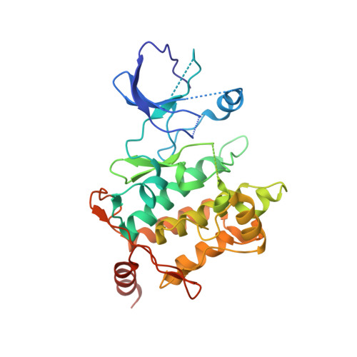
5J7S | pdb_00005j7s

Crystal structure of SM1-71 bound to TAK1-TAB1


Experimental Data Snapshot

  • Method: X-RAY DIFFRACTION
  • Resolution: 2.37 Å
  • R-Value Free: 
    0.240 (Depositor), 0.250 (DCC) 
  • R-Value Work: 
    0.228 (Depositor), 0.230 (DCC) 
  • R-Value Observed: 
    0.229 (Depositor) 

Starting Model: experimental
View more details

wwPDB Validation   3D Report Full Report


Ligand Structure Quality Assessment 

Created with Raphaël 2.3.0Worse 01 BetterLigand structure goodness of fit to experimental dataBest fitted 6H3Click on this verticalbar to view details

This is version 1.4 of the entry. See complete history


Literature

Structure-guided development of covalent TAK1 inhibitors.

Tan, L.Gurbani, D.Weisberg, E.L.Hunter, J.C.Li, L.Jones, D.S.Ficarro, S.B.Mowafy, S.Tam, C.P.Rao, S.Du, G.Griffin, J.D.Sorger, P.K.Marto, J.A.Westover, K.D.Gray, N.S.

(2017) Bioorg Med Chem 25: 838-846

  • DOI: https://doi.org/10.1016/j.bmc.2016.11.035
  • Primary Citation of Related Structures:  
    5E7R, 5J7S, 5J8I, 5J9L, 5JH6, 5JK3

  • PubMed Abstract: 

    TAK1 (transforming growth factor-β-activated kinase 1) is an essential intracellular mediator of cytokine and growth factor signaling and a potential therapeutic target for the treatment of immune diseases and cancer. Herein we report development of a series of 2,4-disubstituted pyrimidine covalent TAK1 inhibitors that target Cys174, a residue immediately adjacent to the 'DFG-motif' of the kinase activation loop. Co-crystal structures of TAK1 with candidate compounds enabled iterative rounds of structure-based design and biological testing to arrive at optimized compounds. Lead compounds such as 2 and 10 showed greater than 10-fold biochemical selectivity for TAK1 over the closely related kinases MEK1 and ERK1 which possess an equivalently positioned cysteine residue. These compounds are smaller, more easily synthesized, and exhibit a different spectrum of kinase selectivity relative to previously reported macrocyclic natural product TAK1 inhibitors such as 5Z-7-oxozeanol.


  • Organizational Affiliation

    Department of Cancer Biology, Dana-Farber Cancer Institute, Boston, MA 02115, USA; Department of Biological Chemistry and Molecular Pharmacology, Harvard Medical School, Boston, MA 02215, USA.


Macromolecules
Find similar proteins by:  (by identity cutoff)  |  3D Structure
Entity ID: 1
MoleculeChains Sequence LengthOrganismDetailsImage
Mitogen-activated protein kinase kinase kinase 7/TGF-beta-activated kinase 1 and MAP3K7-binding protein 1 chimera314Homo sapiensMutation(s): 0 
Gene Names: MAP3K7TAK1TAB1MAP3K7IP1
EC: 2.7.11.25
UniProt & NIH Common Fund Data Resources
Find proteins for Q15750 (Homo sapiens)
Explore Q15750 
Go to UniProtKB:  Q15750
PHAROS:  Q15750
GTEx:  ENSG00000100324 
Find proteins for O43318 (Homo sapiens)
Explore O43318 
Go to UniProtKB:  O43318
PHAROS:  O43318
GTEx:  ENSG00000135341 
Entity Groups  
Sequence Clusters30% Identity50% Identity70% Identity90% Identity95% Identity100% Identity
UniProt GroupsO43318Q15750
Sequence Annotations
Expand
  • Reference Sequence
Small Molecules
Ligands 1 Unique
IDChains Name / Formula / InChI Key2D Diagram3D Interactions
6H3
Query on 6H3

Download Ideal Coordinates CCD File 
B [auth A]N-{2-[(5-chloro-2-{[4-(4-methylpiperazin-1-yl)phenyl]amino}pyrimidin-4-yl)amino]phenyl}propanamide
C24 H28 Cl N7 O
IIMJCIGRKJCCHT-UHFFFAOYSA-N
Binding Affinity Annotations 
IDSourceBinding Affinity
6H3 BindingDB:  5J7S IC50: 1630 (nM) from 1 assay(s)
Experimental Data & Validation

Experimental Data

  • Method: X-RAY DIFFRACTION
  • Resolution: 2.37 Å
  • R-Value Free:  0.240 (Depositor), 0.250 (DCC) 
  • R-Value Work:  0.228 (Depositor), 0.230 (DCC) 
  • R-Value Observed: 0.229 (Depositor) 
Space Group: I 2 2 2
Unit Cell:
Length ( Å )Angle ( ˚ )
a = 58.234α = 90
b = 134.06β = 90
c = 146.814γ = 90
Software Package:
Software NamePurpose
PHENIXrefinement
HKL-3000data reduction
SCALEPACKdata scaling
PHASERphasing

Structure Validation

View Full Validation Report


Ligand Structure Quality Assessment 

Created with Raphaël 2.3.0Worse 01 BetterLigand structure goodness of fit to experimental dataBest fitted 6H3Click on this verticalbar to view details

Entry History & Funding Information

Deposition Data


Funding OrganizationLocationGrant Number
Cancer Prevention and Research Institute of Texas (CPRIT)United StatesCPRIT R1207 and CPRIT RP140233

Revision History  (Full details and data files)

  • Version 1.0: 2017-02-15
    Type: Initial release
  • Version 1.1: 2017-09-27
    Changes: Author supporting evidence
  • Version 1.2: 2020-01-08
    Changes: Author supporting evidence
  • Version 1.3: 2023-09-27
    Changes: Data collection, Database references, Refinement description
  • Version 1.4: 2024-11-06
    Changes: Structure summary