5KXI

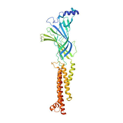
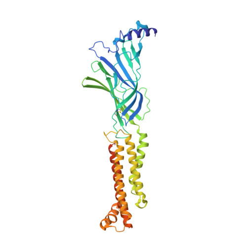
X-ray structure of the human Alpha4Beta2 nicotinic receptor

  • Classification: TRANSPORT PROTEIN
  • Organism(s): Homo sapiens
  • Expression System: Homo sapiens
  • Mutation(s): No 

  • Deposited: 2016-07-20 Released: 2016-09-28 
  • Deposition Author(s): Morales-Perez, C.L., Noviello, C.M., Hibbs, R.E.
  • Funding Organization(s): National Institutes of Health/National Institute of Neurological Disorders and Stroke (NIH/NINDS), Howard Hughes Medical Institute (HHMI), National Institutes of Health/National Institute on Drug Abuse (NIH/NIDA), Welch Foundation, McKnight Scholar Award, Klingenstein-Simons Fellowship Award

Experimental Data Snapshot

  • Method: X-RAY DIFFRACTION
  • Resolution: 3.94 Å
  • R-Value Free: 0.307 
  • R-Value Work: 0.285 
  • R-Value Observed: 0.286 

Starting Model: in silico
View more details

wwPDB Validation   3D Report Full Report


Ligand Structure Quality Assessment 


This is version 1.7 of the entry. See complete history


Literature

X-ray structure of the human alpha 4 beta 2 nicotinic receptor.

Morales-Perez, C.L.Noviello, C.M.Hibbs, R.E.

(2016) Nature 538: 411-415

  • DOI: https://doi.org/10.1038/nature19785
  • Primary Citation of Related Structures:  
    5KXI

  • PubMed Abstract: 

    Nicotinic acetylcholine receptors are ligand-gated ion channels that mediate fast chemical neurotransmission at the neuromuscular junction and have diverse signalling roles in the central nervous system. The nicotinic receptor has been a model system for cell-surface receptors, and specifically for ligand-gated ion channels, for well over a century. In addition to the receptors' prominent roles in the development of the fields of pharmacology and neurobiology, nicotinic receptors are important therapeutic targets for neuromuscular disease, addiction, epilepsy and for neuromuscular blocking agents used during surgery. The overall architecture of the receptor was described in landmark studies of the nicotinic receptor isolated from the electric organ of Torpedo marmorata. Structures of a soluble ligand-binding domain have provided atomic-scale insights into receptor-ligand interactions, while high-resolution structures of other members of the pentameric receptor superfamily provide touchstones for an emerging allosteric gating mechanism. All available high-resolution structures are of homopentameric receptors. However, the vast majority of pentameric receptors (called Cys-loop receptors in eukaryotes) present physiologically are heteromeric. Here we present the X-ray crystallographic structure of the human α4β2 nicotinic receptor, the most abundant nicotinic subtype in the brain. This structure provides insights into the architectural principles governing ligand recognition, heteromer assembly, ion permeation and desensitization in this prototypical receptor class.


  • Organizational Affiliation

    Departments of Neuroscience and Biophysics, University of Texas Southwestern Medical Center, Dallas, Texas 75390, USA.


Macromolecules
Find similar proteins by:  (by identity cutoff)  |  3D Structure
Entity ID: 1
MoleculeChains Sequence LengthOrganismDetailsImage
Neuronal acetylcholine receptor subunit alpha-4
A, D
386Homo sapiensMutation(s): 0 
Gene Names: CHRNA4NACRA4
UniProt & NIH Common Fund Data Resources
Find proteins for P43681 (Homo sapiens)
Go to UniProtKB:  P43681
PHAROS:  P43681
GTEx:  ENSG00000101204 
Glycosylation
Glycosylation Sites: 1Go to GlyGen: P43681-1
Sequence Annotations
Expand
  • Reference Sequence
Find similar proteins by:  (by identity cutoff)  |  3D Structure
Entity ID: 2
MoleculeChains Sequence LengthOrganismDetailsImage
Neuronal acetylcholine receptor subunit beta-2
B, C, E
403Homo sapiensMutation(s): 0 
Gene Names: CHRNB2
UniProt & NIH Common Fund Data Resources
Find proteins for P17787 (Homo sapiens)
Go to UniProtKB:  P17787
PHAROS:  P17787
GTEx:  ENSG00000160716 
Glycosylation
Glycosylation Sites: 1Go to GlyGen: P17787-1
Sequence Annotations
Expand
  • Reference Sequence
Small Molecules
Ligands 3 Unique
IDChains Name / Formula / InChI Key2D Diagram3D Interactions
NAG
Query on NAG

Download Ideal Coordinates CCD File 
F [auth A],
I [auth B],
J [auth C],
K [auth D],
M [auth E]
2-acetamido-2-deoxy-beta-D-glucopyranose
C8 H15 N O6
OVRNDRQMDRJTHS-FMDGEEDCSA-N
NCT
Query on NCT

Download Ideal Coordinates CCD File 
G [auth A],
L [auth D]
(S)-3-(1-METHYLPYRROLIDIN-2-YL)PYRIDINE
C10 H14 N2
SNICXCGAKADSCV-JTQLQIEISA-N
NA
Query on NA

Download Ideal Coordinates CCD File 
H [auth A]SODIUM ION
Na
FKNQFGJONOIPTF-UHFFFAOYSA-N
Binding Affinity Annotations 
IDSourceBinding Affinity
NCT BindingDB:  5KXI Ki: min: 0.05, max: 150 (nM) from 78 assay(s)
IC50: min: 1.2, max: 430 (nM) from 10 assay(s)
EC50: min: 1, max: 1.00e+4 (nM) from 25 assay(s)
Experimental Data & Validation

Experimental Data

  • Method: X-RAY DIFFRACTION
  • Resolution: 3.94 Å
  • R-Value Free: 0.307 
  • R-Value Work: 0.285 
  • R-Value Observed: 0.286 
  • Space Group: P 21 21 21
Unit Cell:
Length ( Å )Angle ( ˚ )
a = 127.099α = 90
b = 132.633β = 90
c = 202.44γ = 90
Software Package:
Software NamePurpose
PHENIXrefinement
HKL-2000data reduction
HKL-2000data scaling
PHENIXphasing

Structure Validation

View Full Validation Report


Ligand Structure Quality Assessment 


Entry History & Funding Information

Deposition Data


Funding OrganizationLocationGrant Number
National Institutes of Health/National Institute of Neurological Disorders and Stroke (NIH/NINDS)United StatesT32 NS069562
Howard Hughes Medical Institute (HHMI)United StatesGilliam Fellowship
National Institutes of Health/National Institute on Drug Abuse (NIH/NIDA)United StatesDA037492
National Institutes of Health/National Institute of Neurological Disorders and Stroke (NIH/NINDS)United StatesNS077983
National Institutes of Health/National Institute of Neurological Disorders and Stroke (NIH/NINDS)United StatesNS095899
Welch FoundationUnited StatesI-1812
McKnight Scholar AwardUnited States--
Klingenstein-Simons Fellowship AwardUnited States--
National Institutes of Health/National Institute on Drug Abuse (NIH/NIDA)United StatesDA042072

Revision History  (Full details and data files)

  • Version 1.0: 2016-09-28
    Type: Initial release
  • Version 1.1: 2016-10-19
    Changes: Database references
  • Version 1.2: 2016-11-02
    Changes: Database references
  • Version 1.3: 2017-09-27
    Changes: Author supporting evidence, Database references, Derived calculations
  • Version 1.4: 2019-11-20
    Changes: Author supporting evidence
  • Version 1.5: 2020-07-29
    Type: Remediation
    Reason: Carbohydrate remediation
    Changes: Data collection, Derived calculations, Structure summary
  • Version 1.6: 2024-04-03
    Changes: Data collection, Database references, Refinement description, Structure summary
  • Version 1.7: 2024-10-16
    Changes: Structure summary