5M5C | pdb_00005m5c

Mechanism of microtubule minus-end recognition and protection by CAMSAP proteins


Experimental Data Snapshot

  • Method: ELECTRON MICROSCOPY
  • Resolution: 4.80 Å
  • Aggregation State: FILAMENT 
  • Reconstruction Method: SINGLE PARTICLE 

wwPDB Validation 3D Report Full Report

Validation slider image for 5M5C

This is version 1.3 of the entry. See complete history

Literature

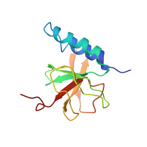
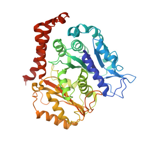
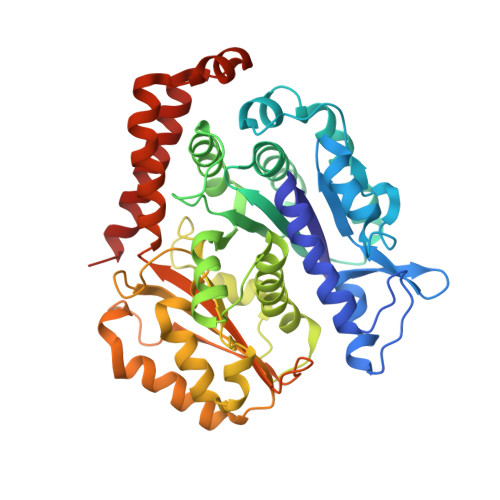
A structural model for microtubule minus-end recognition and protection by CAMSAP proteins.

Atherton, J.Jiang, K.Stangier, M.M.Luo, Y.Hua, S.Houben, K.van Hooff, J.J.E.Joseph, A.P.Scarabelli, G.Grant, B.J.Roberts, A.J.Topf, M.Steinmetz, M.O.Baldus, M.Moores, C.A.Akhmanova, A.

(2017) Nat Struct Mol Biol 24: 931-943

  • DOI: https://doi.org/10.1038/nsmb.3483
  • Primary Citation Related Structures: 
    5LZN, 5M50, 5M54, 5M5C

  • PubMed Abstract: 

    CAMSAP and Patronin family members regulate microtubule minus-end stability and localization and thus organize noncentrosomal microtubule networks, which are essential for cell division, polarization and differentiation. Here, we found that the CAMSAP C-terminal CKK domain is widely present among eukaryotes and autonomously recognizes microtubule minus ends. Through a combination of structural approaches, we uncovered how mammalian CKK binds between two tubulin dimers at the interprotofilament interface on the outer microtubule surface. In vitro reconstitution assays combined with high-resolution fluorescence microscopy and cryo-electron tomography suggested that CKK preferentially associates with the transition zone between curved protofilaments and the regular microtubule lattice. We propose that minus-end-specific features of the interprotofilament interface at this site serve as the basis for CKK's minus-end preference. The steric clash between microtubule-bound CKK and kinesin motors explains how CKK protects microtubule minus ends against kinesin-13-induced depolymerization and thus controls the stability of free microtubule minus ends.


  • Organizational Affiliation
    • Institute of Structural and Molecular Biology, Birkbeck, University of London, London, UK.

Macromolecule Content 

  • Total Structure Weight: 210.14 kDa 
  • Atom Count: 14,608 
  • Modeled Residue Count: 1,826 
  • Deposited Residue Count: 1,846 
  • Unique protein chains: 3

Macromolecules

Find similar proteins by:|  3D Structure
Entity ID: 1
MoleculeChains  Sequence LengthOrganismDetailsImage
Calmodulin-regulated spectrin-associated protein 1A [auth C]118Homo sapiensMutation(s): 1 
Gene Names: CAMSAP1
UniProt & NIH Common Fund Data Resources
Find proteins for Q5T5Y3 (Homo sapiens)
Explore Q5T5Y3 
Go to UniProtKB:  Q5T5Y3
PHAROS:  Q5T5Y3
GTEx:  ENSG00000130559 
Entity Groups
Sequence Clusters30% Identity50% Identity70% Identity90% Identity95% Identity100% Identity
UniProt GroupQ5T5Y3
Sequence Annotations
Expand
Reference Sequence
Find similar proteins by:|  3D Structure
Entity ID: 2
MoleculeChains  Sequence LengthOrganismDetailsImage
Tubulin alpha chainB [auth D],
D [auth A]
438Bos taurusMutation(s): 0 
EC: 3.6.5
UniProt
Find proteins for P81947 (Bos taurus)
Explore P81947 
Go to UniProtKB:  P81947
Entity Groups
Sequence Clusters30% Identity50% Identity70% Identity90% Identity95% Identity100% Identity
UniProt GroupP81947
Sequence Annotations
Expand
Reference Sequence
Find similar proteins by:|  3D Structure
Entity ID: 3
MoleculeChains  Sequence LengthOrganismDetailsImage
Tubulin beta-2B chainC [auth E],
E [auth B]
426Bos taurusMutation(s): 0 
UniProt
Find proteins for Q6B856 (Bos taurus)
Explore Q6B856 
Go to UniProtKB:  Q6B856
Entity Groups
Sequence Clusters30% Identity50% Identity70% Identity90% Identity95% Identity100% Identity
UniProt GroupQ6B856
Sequence Annotations
Expand
Reference Sequence

Small Molecules

Ligands 4 Unique
IDChains Name / Formula / InChI Key2D Diagram3D Interactions
TA1

Query on TA1


Download:Ideal Coordinates CCD File
I [auth E],
M [auth B]
TAXOL
C47 H51 N O14
RCINICONZNJXQF-MZXODVADSA-N
GTP

Query on GTP


Download:Ideal Coordinates CCD File
F [auth D],
J [auth A]
GUANOSINE-5'-TRIPHOSPHATE
C10 H16 N5 O14 P3
XKMLYUALXHKNFT-UUOKFMHZSA-N
GDP

Query on GDP


Download:Ideal Coordinates CCD File
H [auth E],
L [auth B]
GUANOSINE-5'-DIPHOSPHATE
C10 H15 N5 O11 P2
QGWNDRXFNXRZMB-UUOKFMHZSA-N
MG

Query on MG


Download:Ideal Coordinates CCD File
G [auth D],
K [auth A]
MAGNESIUM ION
Mg
JLVVSXFLKOJNIY-UHFFFAOYSA-N

Experimental Data & Validation

Experimental Data

  • Method: ELECTRON MICROSCOPY
  • Resolution: 4.80 Å
  • Aggregation State: FILAMENT 
  • Reconstruction Method: SINGLE PARTICLE 

Structure Validation

View Full Validation Report


Entry History 

Revision History  (Full details and data files)

  • Version 1.0: 2017-10-04
    Type: Initial release
  • Version 1.1: 2017-10-18
    Changes: Database references
  • Version 1.2: 2017-11-15
    Changes: Database references
  • Version 1.3: 2024-05-15
    Changes: Data collection, Database references, Derived calculations