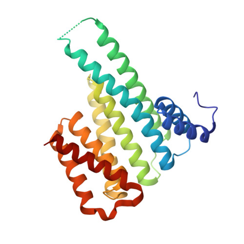
5OEH | pdb_00005oeh

Molecular tweezers modulate 14-3-3 protein-protein interactions.


Experimental Data Snapshot

  • Method: X-RAY DIFFRACTION
  • Resolution: 2.35 Å
  • R-Value Free: 
    0.264 (Depositor), 0.270 (DCC) 
  • R-Value Work: 
    0.202 (Depositor), 0.210 (DCC) 
  • R-Value Observed: 
    0.205 (Depositor) 

Starting Model: experimental
View more details

wwPDB Validation   3D Report Full Report


Ligand Structure Quality Assessment 

Created with Raphaël 2.3.0Worse 01 BetterLigand structure goodness of fit to experimental dataBest fitted 9SZClick on this verticalbar to view details

This is version 1.3 of the entry. See complete history


Literature

Molecular tweezers modulate 14-3-3 protein-protein interactions.

Bier, D.Rose, R.Bravo-Rodriguez, K.Bartel, M.Ramirez-Anguita, J.M.Dutt, S.Wilch, C.Klarner, F.G.Sanchez-Garcia, E.Schrader, T.Ottmann, C.

(2013) Nat Chem 5: 234-239

  • DOI: https://doi.org/10.1038/nchem.1570
  • Primary Citation of Related Structures:  
    5OEG, 5OEH

  • PubMed Abstract: 

    Supramolecular chemistry has recently emerged as a promising way to modulate protein functions, but devising molecules that will interact with a protein in the desired manner is difficult as many competing interactions exist in a biological environment (with solvents, salts or different sites for the target biomolecule). We now show that lysine-specific molecular tweezers bind to a 14-3-3 adapter protein and modulate its interaction with partner proteins. The tweezers inhibit binding between the 14-3-3 protein and two partner proteins--a phosphorylated (C-Raf) protein and an unphosphorylated one (ExoS)--in a concentration-dependent manner. Protein crystallography shows that this effect arises from the binding of the tweezers to a single surface-exposed lysine (Lys214) of the 14-3-3 protein in the proximity of its central channel, which normally binds the partner proteins. A combination of structural analysis and computer simulations provides rules for the tweezers' binding preferences, thus allowing us to predict their influence on this type of protein-protein interactions.


  • Organizational Affiliation

    Chemical Genomics Centre of the Max-Planck-Society, Otto-Hahn-Strasse 15, 44227 Dortmund, Germany.


Macromolecules
Find similar proteins by:  (by identity cutoff)  |  3D Structure
Entity ID: 1
MoleculeChains Sequence LengthOrganismDetailsImage
14-3-3 protein sigma236Homo sapiensMutation(s): 0 
Gene Names: SFNHME1
UniProt & NIH Common Fund Data Resources
Find proteins for P31947 (Homo sapiens)
Explore P31947 
Go to UniProtKB:  P31947
PHAROS:  P31947
GTEx:  ENSG00000175793 
Entity Groups  
Sequence Clusters30% Identity50% Identity70% Identity90% Identity95% Identity100% Identity
UniProt GroupP31947
Sequence Annotations
Expand
  • Reference Sequence
Small Molecules
Ligands 3 Unique
IDChains Name / Formula / InChI Key2D Diagram3D Interactions
9SZ
Query on 9SZ

Download Ideal Coordinates CCD File 
B [auth A](1R,5S,9S,16R,20R,24S,28S,35R)-3,22-Bis(dihydroxyphosphoryloxy)tridecacyclo[22.14.1.15,20.19,16.128,35.02,23.04,21.06,19.08,17.010,15.025,38.027,36.029,34]dotetraconta-2(23),3,6,8(17),10,12,14,18,21,25,27(36),29,31,33,37-pentadecaene
C42 H32 O8 P2
BZESKQAEKFQSPD-FCODNRGNSA-N
GOL
Query on GOL

Download Ideal Coordinates CCD File 
D [auth A]GLYCEROL
C3 H8 O3
PEDCQBHIVMGVHV-UHFFFAOYSA-N
CL
Query on CL

Download Ideal Coordinates CCD File 
C [auth A]CHLORIDE ION
Cl
VEXZGXHMUGYJMC-UHFFFAOYSA-M
Experimental Data & Validation

Experimental Data

  • Method: X-RAY DIFFRACTION
  • Resolution: 2.35 Å
  • R-Value Free:  0.264 (Depositor), 0.270 (DCC) 
  • R-Value Work:  0.202 (Depositor), 0.210 (DCC) 
  • R-Value Observed: 0.205 (Depositor) 
Space Group: C 2 2 2
Unit Cell:
Length ( Å )Angle ( ˚ )
a = 60.4α = 90
b = 157.4β = 90
c = 77.2γ = 90
Software Package:
Software NamePurpose
XSCALEdata scaling
PHASERphasing
REFMACrefinement
PDB_EXTRACTdata extraction
XDSdata reduction

Structure Validation

View Full Validation Report


Ligand Structure Quality Assessment 

Created with Raphaël 2.3.0Worse 01 BetterLigand structure goodness of fit to experimental dataBest fitted 9SZClick on this verticalbar to view details

Entry History 

Deposition Data

Revision History  (Full details and data files)

  • Version 1.0: 2017-07-26
    Type: Initial release
  • Version 1.1: 2020-04-29
    Changes: Derived calculations
  • Version 1.2: 2024-01-10
    Changes: Data collection, Database references, Derived calculations, Structure summary
  • Version 1.3: 2024-01-17
    Changes: Refinement description