


Crystal Structure of a COMT complex
Lerner, C., Jakob-Roetne, R., Groebke-Zbinden, K., Buettelmann, B., Rudolph, M.G.To be published.
Experimental Data Snapshot
Starting Model: other
View more details
Entity ID: 1 | |||||
|---|---|---|---|---|---|
| Molecule | Chains | Sequence Length | Organism | Details | Image |
| Catechol O-methyltransferase | 221 | Rattus norvegicus | Mutation(s): 2 Gene Names: Comt EC: 2.1.1.6 | 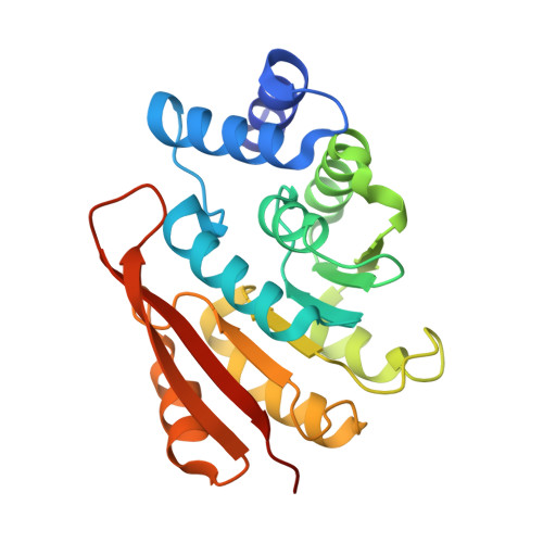 | |
UniProt | |||||
Entity Groups | |||||
| Sequence Clusters | 30% Identity50% Identity70% Identity90% Identity95% Identity100% Identity | ||||
| UniProt Group | P22734 | ||||
Sequence AnnotationsExpand | |||||
Reference Sequence | |||||
| Ligands 7 Unique | |||||
|---|---|---|---|---|---|
| ID | Chains | Name / Formula / InChI Key | 2D Diagram | 3D Interactions | |
| SAH Download:Ideal Coordinates CCD File | C [auth A] | S-ADENOSYL-L-HOMOCYSTEINE C14 H20 N6 O5 S ZJUKTBDSGOFHSH-WFMPWKQPSA-N |  | ||
| 7JF Download:Ideal Coordinates CCD File | B [auth A] | 7-fluoranyl-5,6-bis(4-fluorophenyl)-8-oxidanyl-3~{H}-quinazolin-4-one C20 H11 F3 N2 O2 TXLMFDIODMWHDL-UHFFFAOYSA-N |  | ||
| NHE Download:Ideal Coordinates CCD File | H [auth A], I [auth A] | 2-[N-CYCLOHEXYLAMINO]ETHANE SULFONIC ACID C8 H17 N O3 S MKWKNSIESPFAQN-UHFFFAOYSA-N |  | ||
| DTD Download:Ideal Coordinates CCD File | E [auth A] | DITHIANE DIOL C4 H8 O2 S2 YPGMOWHXEQDBBV-IMJSIDKUSA-N |  | ||
| SO4 Download:Ideal Coordinates CCD File | F [auth A], G [auth A] | SULFATE ION O4 S QAOWNCQODCNURD-UHFFFAOYSA-L |  | ||
| CL Download:Ideal Coordinates CCD File | D [auth A] | CHLORIDE ION Cl VEXZGXHMUGYJMC-UHFFFAOYSA-M |  | ||
| MG Download:Ideal Coordinates CCD File | J [auth A] | MAGNESIUM ION Mg JLVVSXFLKOJNIY-UHFFFAOYSA-N |  | ||
| Length ( Å ) | Angle ( ˚ ) |
|---|---|
| a = 49.711 | α = 90 |
| b = 54.666 | β = 90 |
| c = 81.533 | γ = 90 |
| Software Name | Purpose |
|---|---|
| REFMAC | refinement |
| XSCALE | data scaling |
| PDB_EXTRACT | data extraction |
| XDS | data reduction |
| PHASER | phasing |


