5T0A

Crystal Structure of Heparan Sulfate 6-O-Sulfotransferase with bound PAP and heptasaccharide substrate


Experimental Data Snapshot

  • Method: X-RAY DIFFRACTION
  • Resolution: 1.95 Å
  • R-Value Free: 
    0.213 (Depositor), 0.215 (DCC) 
  • R-Value Work: 
    0.180 (Depositor), 0.181 (DCC) 
  • R-Value Observed: 
    0.181 (Depositor) 

wwPDB Validation   3D Report Full Report


Ligand Structure Quality Assessment 

Created with Raphaël 2.3.0Worse 01 BetterLigand structure goodness of fit to experimental dataBest fitted A3PClick on this verticalbar to view details

This is version 2.1 of the entry. See complete history


Literature

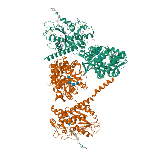
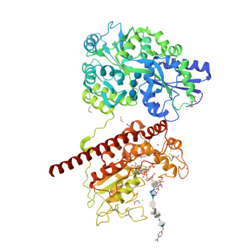
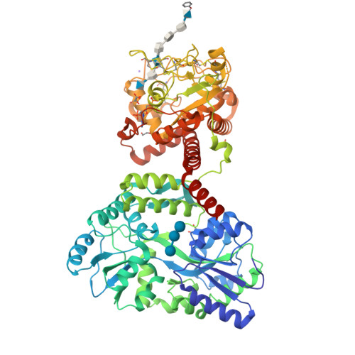
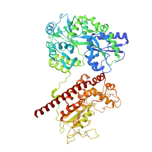
Structure Based Substrate Specificity Analysis of Heparan Sulfate 6-O-Sulfotransferases.

Xu, Y.Moon, A.F.Xu, S.Krahn, J.M.Liu, J.Pedersen, L.C.

(2017) ACS Chem Biol 12: 73-82

  • DOI: https://doi.org/10.1021/acschembio.6b00841
  • Primary Citation of Related Structures:  
    5T03, 5T05, 5T0A

  • PubMed Abstract: 

    Heparan sulfate (HS) is a sulfated polysaccharide exhibiting essential physiological functions. HS 6-O-sulfotransferase (6-OST) transfers a sulfo group to the 6-OH position of glucosamine units to confer a variety of HS biological activities. There are three different isoforms of 6-OST in the human genome. Here, we report crystal structures of the ternary complex of 6-OST with the sulfo donor analog 3'-phosphoadenosine 5'-phosphate and three different oligosaccharide substrates at 1.95 to 2.1 Å resolutions. Structural and mutational analyses reveal amino acid residues that contribute to catalysis and substrate recognition of 6-OST. Unexpectedly, the structures reveal 6-OST engages HS in a completely different orientation than other HS sulfotransferases and sheds light on the basic HS requirements for specificity. These findings also contribute structural information to understand mutations in human 6-OST isoform 1 associated with the human genetic disease idiopathic hypogonadotropic hypogonadism characterized by incomplete or lack of puberty.


  • Organizational Affiliation

    Division of Chemical Biology and Medicinal Chemistry, Eshelman School of Pharmacy, University of North Carolina , Chapel Hill, North Carolina 27599, United States.


Macromolecules
Find similar proteins by:  (by identity cutoff)  |  3D Structure
Entity ID: 1
MoleculeChains Sequence LengthOrganismDetailsImage
maltose binding protein - heparan sulfate 6-O-sulfotransferase isoform 3 fusion protein
A, B
692Escherichia coli O157:H7Danio rerio
This entity is chimeric
Mutation(s): 8 
Gene Names: malEZ5632ECs5017hs6st3
EC: 2.8.2
UniProt
Find proteins for P0AEX9 (Escherichia coli (strain K12))
Explore P0AEX9 
Go to UniProtKB:  P0AEX9
Entity Groups  
Sequence Clusters30% Identity50% Identity70% Identity90% Identity95% Identity100% Identity
UniProt GroupP0AEX9
Sequence Annotations
Expand
  • Reference Sequence
Oligosaccharides

Help

Entity ID: 2
MoleculeChains Length2D Diagram Glycosylation3D Interactions
2-deoxy-2-(sulfoamino)-alpha-D-glucopyranose-(1-4)-beta-D-glucopyranuronic acid-(1-4)-2-deoxy-2-(sulfoamino)-alpha-D-glucopyranose-(1-4)-2-O-sulfo-alpha-L-idopyranuronic acid-(1-4)-2-deoxy-2-(sulfoamino)-alpha-D-glucopyranose-(1-4)-beta-D-glucopyranuronic acid
C
6N/A
Glycosylation Resources
GlyTouCan:  G60898FR
GlyCosmos:  G60898FR
Entity ID: 3
MoleculeChains Length2D Diagram Glycosylation3D Interactions
alpha-D-glucopyranose-(1-4)-alpha-D-glucopyranose-(1-4)-alpha-D-glucopyranose-(1-4)-alpha-D-glucopyranose
D, F
4N/A
Glycosylation Resources
GlyTouCan:  G87171PZ
GlyCosmos:  G87171PZ
GlyGen:  G87171PZ
Entity ID: 4
MoleculeChains Length2D Diagram Glycosylation3D Interactions
beta-D-glucopyranuronic acid-(1-4)-2-deoxy-2-(sulfoamino)-alpha-D-glucopyranose-(1-4)-beta-D-glucopyranuronic acid-(1-4)-2-deoxy-2-(sulfoamino)-alpha-D-glucopyranose-(1-4)-2-O-sulfo-alpha-L-idopyranuronic acid-(1-4)-2-deoxy-2-(sulfoamino)-alpha-D-glucopyranose-(1-4)-beta-D-glucopyranuronic acid
E
7N/A
Glycosylation Resources
GlyTouCan:  G26496DZ
GlyCosmos:  G26496DZ
Small Molecules
Ligands 6 Unique
IDChains Name / Formula / InChI Key2D Diagram3D Interactions
A3P
Query on A3P

Download Ideal Coordinates CCD File 
CA [auth B],
H [auth A]
ADENOSINE-3'-5'-DIPHOSPHATE
C10 H15 N5 O10 P2
WHTCPDAXWFLDIH-KQYNXXCUSA-N
TLA
Query on TLA

Download Ideal Coordinates CCD File 
HA [auth B],
T [auth A],
U [auth A]
L(+)-TARTARIC ACID
C4 H6 O6
FEWJPZIEWOKRBE-JCYAYHJZSA-N
NPO
Query on NPO

Download Ideal Coordinates CCD File 
BA [auth B],
G [auth A]
P-NITROPHENOL
C6 H5 N O3
BTJIUGUIPKRLHP-UHFFFAOYSA-N
EDO
Query on EDO

Download Ideal Coordinates CCD File 
DA [auth B]
EA [auth B]
FA [auth B]
GA [auth B]
I [auth A]
1,2-ETHANEDIOL
C2 H6 O2
LYCAIKOWRPUZTN-UHFFFAOYSA-N
CL
Query on CL

Download Ideal Coordinates CCD File 
AA [auth A],
KA [auth B],
Z [auth A]
CHLORIDE ION
Cl
VEXZGXHMUGYJMC-UHFFFAOYSA-M
NA
Query on NA

Download Ideal Coordinates CCD File 
IA [auth B]
JA [auth B]
V [auth A]
W [auth A]
X [auth A]
SODIUM ION
Na
FKNQFGJONOIPTF-UHFFFAOYSA-N
Biologically Interesting Molecules (External Reference) 1 Unique
Experimental Data & Validation

Experimental Data

  • Method: X-RAY DIFFRACTION
  • Resolution: 1.95 Å
  • R-Value Free:  0.213 (Depositor), 0.215 (DCC) 
  • R-Value Work:  0.180 (Depositor), 0.181 (DCC) 
  • R-Value Observed: 0.181 (Depositor) 
Space Group: P 21 21 21
Unit Cell:
Length ( Å )Angle ( ˚ )
a = 75.775α = 90
b = 128.291β = 90
c = 178.929γ = 90
Software Package:
Software NamePurpose
PHENIXrefinement
HKL-2000data reduction
HKL-2000data scaling
PHASERphasing

Structure Validation

View Full Validation Report


Ligand Structure Quality Assessment 

Created with Raphaël 2.3.0Worse 01 BetterLigand structure goodness of fit to experimental dataBest fitted A3PClick on this verticalbar to view details

Entry History & Funding Information

Deposition Data


Funding OrganizationLocationGrant Number
National Institutes of Health/National Institute of General Medical Sciences (NIH/NIGMS)United StatesGM102137
National Institutes of Health/National Heart, Lung, and Blood Institute (NIH/NHLBI)United StatesHL094463

Revision History  (Full details and data files)

  • Version 1.0: 2017-02-01
    Type: Initial release
  • Version 1.1: 2017-09-20
    Changes: Author supporting evidence
  • Version 1.2: 2019-12-04
    Changes: Author supporting evidence, Data collection
  • Version 2.0: 2020-07-29
    Type: Remediation
    Reason: Carbohydrate remediation
    Changes: Advisory, Atomic model, Data collection, Derived calculations, Structure summary
  • Version 2.1: 2024-11-06
    Changes: Data collection, Database references, Derived calculations, Structure summary