5U3F | pdb_00005u3f

Structure of Mycobacterium tuberculosis IlvE, a branched-chain amino acid transaminase, in complex with D-cycloserine derivative


Experimental Data Snapshot

  • Method: X-RAY DIFFRACTION
  • Resolution: 1.70 Å
  • R-Value Free: 
    0.210 (Depositor), 0.210 (DCC) 
  • R-Value Work: 
    0.176 (Depositor), 0.180 (DCC) 
  • R-Value Observed: 
    0.178 (Depositor) 

Starting Model: experimental
View more details

wwPDB Validation   3D Report Full Report


Ligand Structure Quality Assessment 

Created with Raphaël 2.3.0Worse 01 BetterLigand structure goodness of fit to experimental dataBest fitted 7TSClick on this verticalbar to view details

This is version 1.5 of the entry. See complete history


Literature

Mechanism-Based Inhibition of the Mycobacterium tuberculosis Branched-Chain Aminotransferase by d- and l-Cycloserine.

Amorim Franco, T.M.Favrot, L.Vergnolle, O.Blanchard, J.S.

(2017) ACS Chem Biol 12: 1235-1244

  • DOI: https://doi.org/10.1021/acschembio.7b00142
  • Primary Citation of Related Structures:  
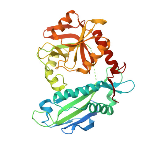
    5U3F

  • PubMed Abstract: 

    The branched-chain aminotransferase is a pyridoxal 5'-phosphate (PLP)-dependent enzyme responsible for the final step in the biosynthesis of all three branched-chain amino acids, l-leucine, l-isoleucine, and l-valine, in bacteria. We have investigated the mechanism of inactivation of the branched-chain aminotransferase from Mycobacterium tuberculosis (MtIlvE) by d- and l-cycloserine. d-Cycloserine is currently used only in the treatment of multidrug-drug-resistant tuberculosis. Our results show a time- and concentration-dependent inactivation of MtIlvE by both isomers, with l-cycloserine being a 40-fold better inhibitor of the enzyme. Minimum inhibitory concentration (MIC) studies revealed that l-cycloserine is a 10-fold better inhibitor of Mycobacterium tuberculosis growth than d-cycloserine. In addition, we have crystallized the MtIlvE-d-cycloserine inhibited enzyme, determining the structure to 1.7 Å. The structure of the covalent d-cycloserine-PMP adduct bound to MtIlvE reveals that the d-cycloserine ring is planar and aromatic, as previously observed for other enzyme systems. Mass spectrometry reveals that both the d-cycloserine- and l-cycloserine-PMP complexes have the same mass, and are likely to be the same aromatized, isoxazole product. However, the kinetics of formation of the MtIlvE d-cycloserine-PMP and MtIlvE l-cycloserine-PMP adducts are quite different. While the kinetics of the formation of the MtIlvE d-cycloserine-PMP complex can be fit to a single exponential, the formation of the MtIlvE l-cycloserine-PMP complex occurs in two steps. We propose a chemical mechanism for the inactivation of d- and l-cycloserine which suggests a stereochemically determined structural role for the differing kinetics of inactivation. These results demonstrate that the mechanism of action of d-cycloserine's activity against M. tuberculosis may be more complicated than previously thought and that d-cycloserine may compromise the in vivo activity of multiple PLP-dependent enzymes, including MtIlvE.


  • Organizational Affiliation

    Department of Biochemistry, Albert Einstein College of Medicine , 1300 Morris Park Avenue, Bronx, New York 10461, United States.


Macromolecules
Find similar proteins by:  (by identity cutoff)  |  3D Structure
Entity ID: 1
MoleculeChains Sequence LengthOrganismDetailsImage
Branched-chain-amino-acid aminotransferase
A, B
368Mycobacterium tuberculosis H37RvMutation(s): 0 
Gene Names: ilvERv2210cMTCY190.21c
EC: 2.6.1.42
UniProt
Find proteins for P9WQ75 (Mycobacterium tuberculosis (strain ATCC 25618 / H37Rv))
Explore P9WQ75 
Go to UniProtKB:  P9WQ75
Entity Groups  
Sequence Clusters30% Identity50% Identity70% Identity90% Identity95% Identity100% Identity
UniProt GroupP9WQ75
Sequence Annotations
Expand
  • Reference Sequence
Small Molecules
Ligands 1 Unique
IDChains Name / Formula / InChI Key2D Diagram3D Interactions
7TS
Query on 7TS

Download Ideal Coordinates CCD File 
C [auth A],
D [auth B]
(5-hydroxy-6-methyl-4-{[(3-oxo-2,3-dihydro-1,2-oxazol-4-yl)amino]methyl}pyridin-3-yl)methyl dihydrogen phosphate
C11 H14 N3 O7 P
PXWFNGNWQUPGPJ-UHFFFAOYSA-N
Experimental Data & Validation

Experimental Data

  • Method: X-RAY DIFFRACTION
  • Resolution: 1.70 Å
  • R-Value Free:  0.210 (Depositor), 0.210 (DCC) 
  • R-Value Work:  0.176 (Depositor), 0.180 (DCC) 
  • R-Value Observed: 0.178 (Depositor) 
Space Group: P 21 21 21
Unit Cell:
Length ( Å )Angle ( ˚ )
a = 91.892α = 90
b = 80.464β = 90
c = 81.25γ = 90
Software Package:
Software NamePurpose
PHENIXrefinement
XDSdata reduction
XDSdata scaling
PHASERphasing

Structure Validation

View Full Validation Report


Ligand Structure Quality Assessment 

Created with Raphaël 2.3.0Worse 01 BetterLigand structure goodness of fit to experimental dataBest fitted 7TSClick on this verticalbar to view details

Entry History & Funding Information

Deposition Data


Funding OrganizationLocationGrant Number
National Institutes of Health/National Institute Of Allergy and Infectious Diseases (NIH/NIAID)United StatesAI060899
Science Without Boarders fellowship CAPES Coordenacao de Aperfeicoamento de Pessoal de Nivel SuperiorBrazil--

Revision History  (Full details and data files)

  • Version 1.0: 2017-03-22
    Type: Initial release
  • Version 1.1: 2017-05-31
    Changes: Database references
  • Version 1.2: 2017-09-20
    Changes: Author supporting evidence
  • Version 1.3: 2019-12-11
    Changes: Author supporting evidence
  • Version 1.4: 2023-10-04
    Changes: Data collection, Database references, Refinement description
  • Version 1.5: 2024-10-30
    Changes: Structure summary