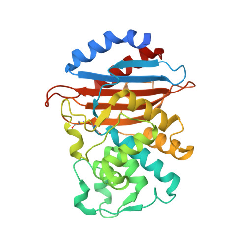
5UJ3

Crystal structure of the KPC-2 beta-lactamase complexed with hydrolyzed cefotaxime


Experimental Data Snapshot

  • Method: X-RAY DIFFRACTION
  • Resolution: 1.45 Å
  • R-Value Free: 0.212 
  • R-Value Work: 0.169 
  • R-Value Observed: 0.171 

wwPDB Validation   3D Report Full Report


Ligand Structure Quality Assessment 


This is version 1.4 of the entry. See complete history


Literature

Molecular Basis of Substrate Recognition and Product Release by the Klebsiella pneumoniae Carbapenemase (KPC-2).

Pemberton, O.A.Zhang, X.Chen, Y.

(2017) J Med Chem 60: 3525-3530

  • DOI: https://doi.org/10.1021/acs.jmedchem.7b00158
  • Primary Citation of Related Structures:  
    5UJ3, 5UJ4, 5UL8

  • PubMed Abstract: 

    Carbapenem-resistant Enterobacteriaceae are resistant to most β-lactam antibiotics due to the production of the Klebsiella pneumoniae carbapenemase (KPC-2) class A β-lactamase. Here, we present the first product complex crystal structures of KPC-2 with β-lactam antibiotics containing hydrolyzed cefotaxime and faropenem. They provide experimental insights into substrate recognition by KPC-2 and its unique cephalosporinase/carbapenemase activity. These structures also represent the first product complexes for a wild-type serine β-lactamase, elucidating the product release mechanism of these enzymes in general.


  • Organizational Affiliation

    Department of Molecular Medicine, University of South Florida , 12901 Bruce B. Downs Boulevard, Tampa, Florida 33612, United States.


Macromolecules
Find similar proteins by:  (by identity cutoff)  |  3D Structure
Entity ID: 1
MoleculeChains Sequence LengthOrganismDetailsImage
Carbapenem-hydrolyzing beta-lactamase KPC290Klebsiella pneumoniaeMutation(s): 0 
Gene Names: blakpckpc1
EC: 3.5.2.6
UniProt
Find proteins for Q9F663 (Klebsiella pneumoniae)
Explore Q9F663 
Go to UniProtKB:  Q9F663
Entity Groups  
Sequence Clusters30% Identity50% Identity70% Identity90% Identity95% Identity100% Identity
UniProt GroupQ9F663
Sequence Annotations
Expand
  • Reference Sequence
Small Molecules
Ligands 2 Unique
IDChains Name / Formula / InChI Key2D Diagram3D Interactions
CE4
Query on CE4

Download Ideal Coordinates CCD File 
B [auth A],
C [auth A]
(2R)-2-[(R)-{[(2Z)-2-(2-amino-1,3-thiazol-4-yl)-2-(methoxyimino)acetyl]amino}(carboxy)methyl]-5-methylidene-5,6-dihydro -2H-1,3-thiazine-4-carboxylic acid
C14 H15 N5 O6 S2
ZRDXEERHKBZJMY-KTFFUYHOSA-N
GOL
Query on GOL

Download Ideal Coordinates CCD File 
D [auth A]GLYCEROL
C3 H8 O3
PEDCQBHIVMGVHV-UHFFFAOYSA-N
Experimental Data & Validation

Experimental Data

  • Method: X-RAY DIFFRACTION
  • Resolution: 1.45 Å
  • R-Value Free: 0.212 
  • R-Value Work: 0.169 
  • R-Value Observed: 0.171 
  • Space Group: P 2 21 21
Unit Cell:
Length ( Å )Angle ( ˚ )
a = 56.93α = 90
b = 58.78β = 90
c = 76.84γ = 90
Software Package:
Software NamePurpose
PHENIXrefinement
SCALAdata scaling
PHASERphasing
iMOSFLMdata reduction
MOSFLMdata reduction

Structure Validation

View Full Validation Report


Ligand Structure Quality Assessment 


Entry History & Funding Information

Deposition Data


Funding OrganizationLocationGrant Number
National Institutes of Health/National Institute Of Allergy and Infectious Diseases (NIH/NIAID)United States4R01AI103158-04

Revision History  (Full details and data files)

  • Version 1.0: 2017-04-26
    Type: Initial release
  • Version 1.1: 2017-05-10
    Changes: Database references
  • Version 1.2: 2017-09-20
    Changes: Author supporting evidence
  • Version 1.3: 2019-12-11
    Changes: Author supporting evidence
  • Version 1.4: 2024-10-16
    Changes: Data collection, Database references, Derived calculations, Structure summary