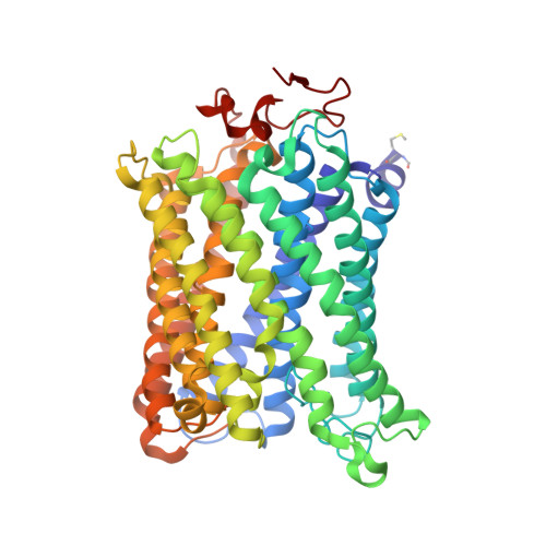
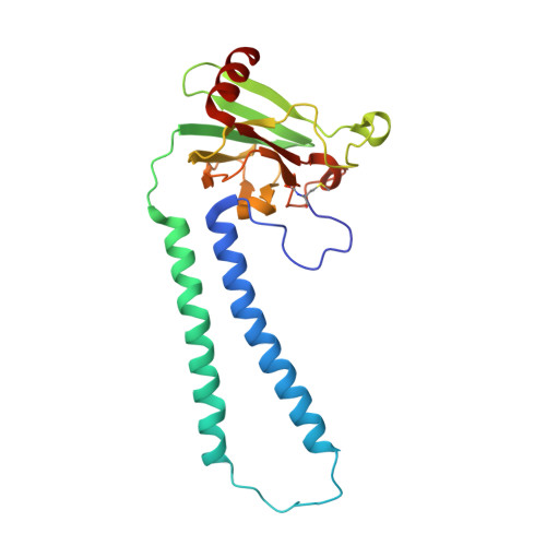
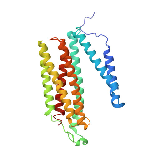
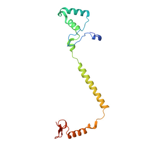
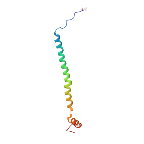
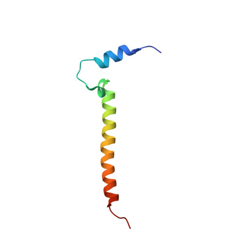
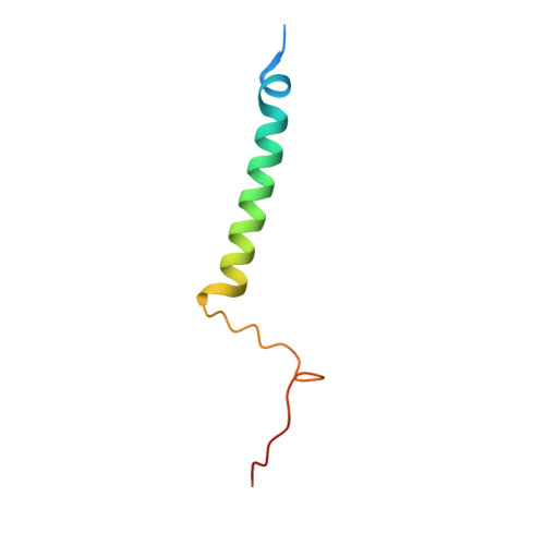
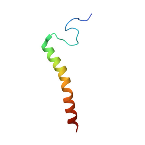
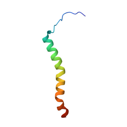
5XDQ

Bovine heart cytochrome c oxidase in the fully oxidized state with pH 7.3 at 1.77 angstrom resolution


Experimental Data Snapshot

  • Method: X-RAY DIFFRACTION
  • Resolution: 1.77 Å
  • R-Value Free: 0.190 
  • R-Value Work: 0.164 
  • R-Value Observed: 0.165 

Starting Model: experimental
View more details

wwPDB Validation   3D Report Full Report


Ligand Structure Quality Assessment 


This is version 1.2 of the entry. See complete history


Literature

Structure of bovine cytochrome c oxidase crystallized at a neutral pH using a fluorinated detergent.

Luo, F.Shinzawa-Itoh, K.Hagimoto, K.Shimada, A.Shimada, S.Yamashita, E.Yoshikawa, S.Tsukihara, T.

(2017) Acta Crystallogr F Struct Biol Commun 73: 416-422

  • DOI: https://doi.org/10.1107/S2053230X17008834
  • Primary Citation of Related Structures:  
    5XDQ

  • PubMed Abstract: 

    Cytochrome c oxidase (CcO) couples proton pumping to O 2 reduction. Its enzymatic activity depends sensitively on pH over a wide range. However, owing to difficulty in crystallizing this protein, X-ray structure analyses of bovine CcO aimed at understanding its reaction mechanism have been conducted using crystals prepared at pH 5.7, which is significantly lower than that in the cell. Here, oxidized CcO at pH 7.3 was crystallized using a fluorinated octyl-maltoside derivative, and the structure was determined at 1.77 Å resolution. No structural differences between crystals obtained at the neutral pH and the acidic pH were detected within the molecules. On the other hand, some differences in intermolecular interactions were detected between the two types of crystal. The influence of pH on the molecular surface is likely to contribute to the pH dependency of the aerobic oxidation of ferrocytochrome c.


  • Organizational Affiliation

    Picobiology Institute, Graduate School of Life Science, University of Hyogo, 3-2-1 Koto, Kamigori-cho, Ako-gun, Hyogo 678-1297, Japan.


Macromolecules
Find similar proteins by:  (by identity cutoff)  |  3D Structure
Entity ID: 1
MoleculeChains Sequence LengthOrganismDetailsImage
Cytochrome c oxidase subunit 1
A, N
514Bos taurusMutation(s): 0 
EC: 1.9.3.1
UniProt
Find proteins for P00396 (Bos taurus)
Go to UniProtKB:  P00396
Sequence Annotations
Expand
  • Reference Sequence
Find similar proteins by:  (by identity cutoff)  |  3D Structure
Entity ID: 2
MoleculeChains Sequence LengthOrganismDetailsImage
Cytochrome c oxidase subunit 2
B, O
227Bos taurusMutation(s): 0 
UniProt
Find proteins for P68530 (Bos taurus)
Go to UniProtKB:  P68530
Sequence Annotations
Expand
  • Reference Sequence
Find similar proteins by:  (by identity cutoff)  |  3D Structure
Entity ID: 3
MoleculeChains Sequence LengthOrganismDetailsImage
Cytochrome c oxidase subunit 3
C, P
261Bos taurusMutation(s): 0 
UniProt
Find proteins for P00415 (Bos taurus)
Go to UniProtKB:  P00415
Sequence Annotations
Expand
  • Reference Sequence
Find similar proteins by:  (by identity cutoff)  |  3D Structure
Entity ID: 4
MoleculeChains Sequence LengthOrganismDetailsImage
Cytochrome c oxidase subunit 4 isoform 1, mitochondrial
D, Q
147Bos taurusMutation(s): 0 
UniProt
Find proteins for P00423 (Bos taurus)
Go to UniProtKB:  P00423
Sequence Annotations
Expand
  • Reference Sequence
Find similar proteins by:  (by identity cutoff)  |  3D Structure
Entity ID: 5
MoleculeChains Sequence LengthOrganismDetailsImage
Cytochrome c oxidase subunit 5A, mitochondrial
E, R
109Bos taurusMutation(s): 0 
UniProt
Find proteins for P00426 (Bos taurus)
Go to UniProtKB:  P00426
Sequence Annotations
Expand
  • Reference Sequence
Find similar proteins by:  (by identity cutoff)  |  3D Structure
Entity ID: 6
MoleculeChains Sequence LengthOrganismDetailsImage
Cytochrome c oxidase subunit 5B, mitochondrial
F, S
94Bos taurusMutation(s): 0 
UniProt
Find proteins for P00428 (Bos taurus)
Go to UniProtKB:  P00428
Sequence Annotations
Expand
  • Reference Sequence
Find similar proteins by:  (by identity cutoff)  |  3D Structure
Entity ID: 7
MoleculeChains Sequence LengthOrganismDetailsImage
Cytochrome c oxidase subunit 6A2, mitochondrial
G, T
85Bos taurusMutation(s): 0 
UniProt
Find proteins for P07471 (Bos taurus)
Go to UniProtKB:  P07471
Sequence Annotations
Expand
  • Reference Sequence
Find similar proteins by:  (by identity cutoff)  |  3D Structure
Entity ID: 8
MoleculeChains Sequence LengthOrganismDetailsImage
Cytochrome c oxidase subunit 6B1
H, U
85Bos taurusMutation(s): 0 
UniProt
Find proteins for P00429 (Bos taurus)
Go to UniProtKB:  P00429
Sequence Annotations
Expand
  • Reference Sequence
Find similar proteins by:  (by identity cutoff)  |  3D Structure
Entity ID: 9
MoleculeChains Sequence LengthOrganismDetailsImage
Cytochrome c oxidase subunit 6C
I, V
73Bos taurusMutation(s): 0 
UniProt
Find proteins for P04038 (Bos taurus)
Go to UniProtKB:  P04038
Sequence Annotations
Expand
  • Reference Sequence
Find similar proteins by:  (by identity cutoff)  |  3D Structure
Entity ID: 10
MoleculeChains Sequence LengthOrganismDetailsImage
Cytochrome c oxidase subunit 7A1, mitochondrial
J, W
59Bos taurusMutation(s): 0 
UniProt
Find proteins for P07470 (Bos taurus)
Go to UniProtKB:  P07470
Sequence Annotations
Expand
  • Reference Sequence
Find similar proteins by:  (by identity cutoff)  |  3D Structure
Entity ID: 11
MoleculeChains Sequence LengthOrganismDetailsImage
Cytochrome c oxidase subunit 7B, mitochondrial
K, X
56Bos taurusMutation(s): 0 
UniProt
Find proteins for P13183 (Bos taurus)
Go to UniProtKB:  P13183
Sequence Annotations
Expand
  • Reference Sequence
Find similar proteins by:  (by identity cutoff)  |  3D Structure
Entity ID: 12
MoleculeChains Sequence LengthOrganismDetailsImage
Cytochrome c oxidase subunit 7C, mitochondrial
L, Y
47Bos taurusMutation(s): 0 
UniProt
Find proteins for P00430 (Bos taurus)
Go to UniProtKB:  P00430
Sequence Annotations
Expand
  • Reference Sequence
Find similar proteins by:  (by identity cutoff)  |  3D Structure
Entity ID: 13
MoleculeChains Sequence LengthOrganismDetailsImage
Cytochrome c oxidase subunit 8B, mitochondrial
M, Z
46Bos taurusMutation(s): 0 
UniProt
Find proteins for P10175 (Bos taurus)
Go to UniProtKB:  P10175
Sequence Annotations
Expand
  • Reference Sequence
Small Molecules
Ligands 15 Unique
IDChains Name / Formula / InChI Key2D Diagram3D Interactions
CDL
Query on CDL

Download Ideal Coordinates CCD File 
AE [auth T],
FD [auth P],
IB [auth C],
UB [auth G]
CARDIOLIPIN
C81 H156 O17 P2
XVTUQDWPJJBEHJ-KZCWQMDCSA-L
TGL
Query on TGL

Download Ideal Coordinates CCD File 
BC [auth L]
OC [auth N]
OD [auth Q]
PB [auth D]
TA [auth B]
BC [auth L],
OC [auth N],
OD [auth Q],
PB [auth D],
TA [auth B],
XC [auth O]
TRISTEAROYLGLYCEROL
C57 H110 O6
DCXXMTOCNZCJGO-UHFFFAOYSA-N
HEA
Query on HEA

Download Ideal Coordinates CCD File 
AA [auth A],
BA [auth A],
JC [auth N],
KC [auth N]
HEME-A
C49 H56 Fe N4 O6
ZGGYGTCPXNDTRV-PRYGPKJJSA-L
PEK
Query on PEK

Download Ideal Coordinates CCD File 
DD [auth P]
EB [auth C]
FB [auth C]
WB [auth G]
YD [auth T]
DD [auth P],
EB [auth C],
FB [auth C],
WB [auth G],
YD [auth T],
ZD [auth T]
(1S)-2-{[(2-AMINOETHOXY)(HYDROXY)PHOSPHORYL]OXY}-1-[(STEAROYLOXY)METHYL]ETHYL (5E,8E,11E,14E)-ICOSA-5,8,11,14-TETRAENOATE
C43 H78 N O8 P
ANRKEHNWXKCXDB-BHFWLYLHSA-N
PSC
Query on PSC

Download Ideal Coordinates CCD File 
VA [auth B],
ZC [auth O]
(7R,17E,20E)-4-HYDROXY-N,N,N-TRIMETHYL-9-OXO-7-[(PALMITOYLOXY)METHYL]-3,5,8-TRIOXA-4-PHOSPHAHEXACOSA-17,20-DIEN-1-AMINIUM 4-OXIDE
C42 H81 N O8 P
JLPULHDHAOZNQI-AUSZDXHESA-O
PGV
Query on PGV

Download Ideal Coordinates CCD File 
DE [auth U]
ED [auth P]
FA [auth A]
GB [auth C]
HA [auth A]
DE [auth U],
ED [auth P],
FA [auth A],
GB [auth C],
HA [auth A],
HB [auth C],
PC [auth N],
RC [auth N]
(1R)-2-{[{[(2S)-2,3-DIHYDROXYPROPYL]OXY}(HYDROXY)PHOSPHORYL]OXY}-1-[(PALMITOYLOXY)METHYL]ETHYL (11E)-OCTADEC-11-ENOATE
C40 H77 O10 P
ADYWCMPUNIVOEA-GPJPVTGXSA-N
DMU
Query on DMU

Download Ideal Coordinates CCD File 
GC [auth M]
HE [auth W]
JE [auth Z]
NB [auth C]
ND [auth P]
GC [auth M],
HE [auth W],
JE [auth Z],
NB [auth C],
ND [auth P],
OB [auth C]
DECYL-BETA-D-MALTOPYRANOSIDE
C22 H42 O11
WOQQAWHSKSSAGF-WXFJLFHKSA-N
CHD
Query on CHD

Download Ideal Coordinates CCD File 
BD [auth P]
CB [auth C]
EE [auth W]
GD [auth P]
JB [auth C]
BD [auth P],
CB [auth C],
EE [auth W],
GD [auth P],
JB [auth C],
VB [auth G],
WA [auth B],
ZB [auth J]
CHOLIC ACID
C24 H40 O5
BHQCQFFYRZLCQQ-OELDTZBJSA-N
CUA
Query on CUA

Download Ideal Coordinates CCD File 
UA [auth B],
YC [auth O]
DINUCLEAR COPPER ION
Cu2
ALKZAGKDWUSJED-UHFFFAOYSA-N
ZN
Query on ZN

Download Ideal Coordinates CCD File 
RB [auth F],
UD [auth S]
ZINC ION
Zn
PTFCDOFLOPIGGS-UHFFFAOYSA-N
CU
Query on CU

Download Ideal Coordinates CCD File 
CA [auth A],
LC [auth N]
COPPER (II) ION
Cu
JPVYNHNXODAKFH-UHFFFAOYSA-N
EDO
Query on EDO

Download Ideal Coordinates CCD File 
AB [auth B]
AC [auth J]
AD [auth O]
BB [auth B]
BE [auth T]
AB [auth B],
AC [auth J],
AD [auth O],
BB [auth B],
BE [auth T],
CC [auth L],
CE [auth T],
DC [auth L],
EC [auth L],
FC [auth L],
FE [auth W],
GE [auth W],
HC [auth M],
HD [auth P],
IA [auth A],
IC [auth M],
ID [auth P],
IE [auth Y],
JA [auth A],
JD [auth P],
KA [auth A],
KB [auth C],
KD [auth P],
LA [auth A],
LB [auth C],
LD [auth P],
MA [auth A],
MB [auth C],
MD [auth P],
NA [auth A],
OA [auth A],
PA [auth A],
PD [auth Q],
QA [auth A],
QB [auth E],
QD [auth R],
RA [auth A],
RD [auth R],
SA [auth A],
SB [auth F],
SC [auth N],
SD [auth R],
TB [auth F],
TC [auth N],
TD [auth R],
UC [auth N],
VC [auth N],
VD [auth S],
WC [auth N],
WD [auth S],
XA [auth B],
XB [auth G],
XD [auth S],
YA [auth B],
YB [auth H],
ZA [auth B]
1,2-ETHANEDIOL
C2 H6 O2
LYCAIKOWRPUZTN-UHFFFAOYSA-N
PER
Query on PER

Download Ideal Coordinates CCD File 
GA [auth A],
QC [auth N]
PEROXIDE ION
O2
ANAIPYUSIMHBEL-UHFFFAOYSA-N
MG
Query on MG

Download Ideal Coordinates CCD File 
DA [auth A],
MC [auth N]
MAGNESIUM ION
Mg
JLVVSXFLKOJNIY-UHFFFAOYSA-N
NA
Query on NA

Download Ideal Coordinates CCD File 
CD [auth P],
DB [auth C],
EA [auth A],
NC [auth N]
SODIUM ION
Na
FKNQFGJONOIPTF-UHFFFAOYSA-N
Modified Residues  3 Unique
IDChains TypeFormula2D DiagramParent
FME
Query on FME
A, N
L-PEPTIDE LINKINGC6 H11 N O3 SMET
TPO
Query on TPO
G, T
L-PEPTIDE LINKINGC4 H10 N O6 PTHR
SAC
Query on SAC
I, V
L-PEPTIDE LINKINGC5 H9 N O4SER
Experimental Data & Validation

Experimental Data

  • Method: X-RAY DIFFRACTION
  • Resolution: 1.77 Å
  • R-Value Free: 0.190 
  • R-Value Work: 0.164 
  • R-Value Observed: 0.165 
  • Space Group: P 21 21 21
Unit Cell:
Length ( Å )Angle ( ˚ )
a = 183.307α = 90
b = 205.904β = 90
c = 177.526γ = 90
Software Package:
Software NamePurpose
REFMACrefinement
SCALEPACKdata scaling
PDB_EXTRACTdata extraction
DENZOdata reduction
MOLREPphasing

Structure Validation

View Full Validation Report


Ligand Structure Quality Assessment 


Entry History & Funding Information

Deposition Data


Funding OrganizationLocationGrant Number
JST CRESTJapanJPMJCR12M3

Revision History  (Full details and data files)

  • Version 1.0: 2017-07-12
    Type: Initial release
  • Version 1.1: 2018-08-15
    Changes: Data collection, Database references
  • Version 1.2: 2023-11-22
    Changes: Data collection, Database references, Derived calculations, Refinement description, Structure summary