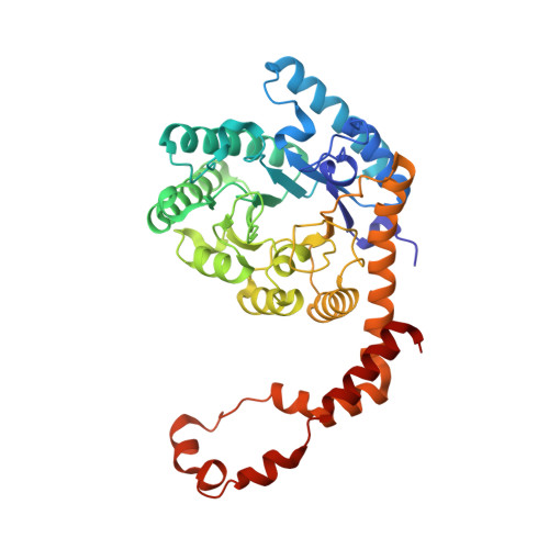
Crystal structure of glucose isomerase in complex with xylitol inhibitor in one metal binding mode
Bae, J.E., Kim, I.J., Nam, K.H.(2017) Biochem Biophys Res Commun 493: 666-670
- PubMed: 28865958
- DOI: https://doi.org/10.1016/j.bbrc.2017.08.134
- Primary Citation of Related Structures:
5Y4I, 5Y4J - PubMed Abstract:
Glucose isomerase (GI) is an intramolecular oxidoreductase that interconverts aldoses and ketoses. These characteristics are widely used in the food, detergent, and pharmaceutical industries. In order to obtain an efficient GI, identification of novel GI genes and substrate binding/inhibition have been studied. Xylitol is a well-known inhibitor of GI. In Streptomyces rubiginosus, two crystal structures have been reported for GI in complex with xylitol inhibitor. However, a structural comparison showed that xylitol can have variable conformation at the substrate binding site, e.g., a nonspecific binding mode. In this study, we report the crystal structure of S. rubiginosus GI in a complex with xylitol and glycerol. Our crystal structure showed one metal binding mode in GI, which we presumed to represent the inactive form of the GI. The metal ion was found only at the M1 site, which was involved in substrate binding, and was not present at the M2 site, which was involved in catalytic function. The O 2 and O 4 atoms of xylitol molecules contributed to the stable octahedral coordination of the metal in M1. Although there was no metal at the M2 site, no large conformational change was observed for the conserved residues coordinating M2. Our structural analysis showed that the metal at the M2 site was not important when a xylitol inhibitor was bound to the M1 site in GI. Thus, these findings provided important information for elucidation or engineering of GI functions.
- Pohang Accelerator Laboratory, POSTECH, Pohang, 35398, Republic of Korea; School of Life Sciences, Kyungpook National University, Daegu, 41566, Republic of Korea.
Organizational Affiliation:


