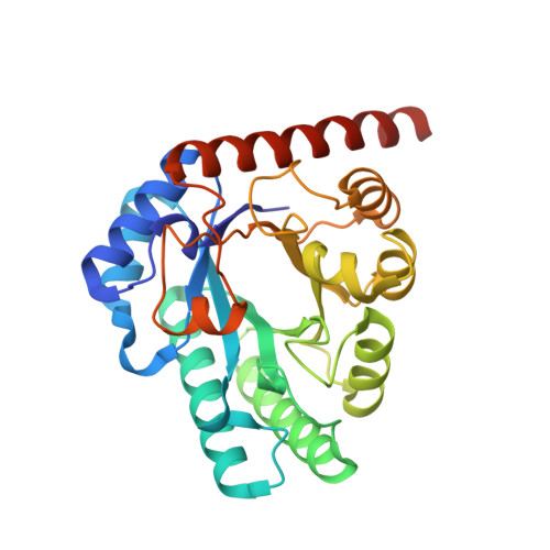
X-ray structure of Arthrobacter globiformis M30 ketose 3-epimerase for the production of D-allulose from D-fructose.
Yoshida, H., Yoshihara, A., Gullapalli, P.K., Ohtani, K., Akimitsu, K., Izumori, K., Kamitori, S.(2018) Acta Crystallogr F Struct Biol Commun 74: 669-676
- PubMed: 30279320
- DOI: https://doi.org/10.1107/S2053230X18011706
- Primary Citation of Related Structures:
5ZFS - PubMed Abstract:
The X-ray structure of ketose 3-epimerase from Arthrobacter globiformis M30, which was previously reported to be a D-allulose 3-epimerase (AgD-AE), was determined at 1.96 Å resolution. The crystal belonged to the hexagonal space group P6 5 22, with unit-cell parameters a = b = 103.98, c = 256.53 Å. The structure was solved by molecular replacement using the structure of Mesorhizobium loti L-ribulose 3-epimerase (MlL-RE), which has 41% sequence identity, as a search model. A hexagonal crystal contained two molecules in the asymmetric unit, and AgD-AE formed a homotetramer with twofold symmetry. The overall structure of AgD-AE was more similar to that of MlL-RE than to the known structures of D-psicose (alternative name D-allulose) 3-epimerases (D-PEs or D-AEs), although AgD-AE and MlL-RE have different substrate specificities. Both AgD-AE and MlL-RE have long helices in the C-terminal region that would contribute to the stability of the homotetramer. AgD-AE showed higher enzymatic activity for L-ribulose than D-allulose; however, AgD-AE is stable and is a unique useful enzyme for the production of D-allulose from D-fructose.
- Life Science Research Center and Faculty of Medicine, Kagawa University, 1750-1 Ikenobe, Miki-cho, Kita-gun, Kagawa 761-0793, Japan.
Organizational Affiliation:


