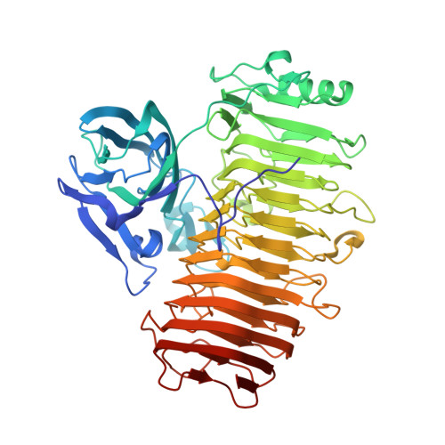
5ZRU

Crystal structure of Agl-KA catalytic domain


Experimental Data Snapshot

  • Method: X-RAY DIFFRACTION
  • Resolution: 1.83 Å
  • R-Value Free: 0.171 
  • R-Value Work: 0.141 
  • R-Value Observed: 0.143 

wwPDB Validation   3D Report Full Report


This is version 1.2 of the entry. See complete history


Literature

Crystal structure of the catalytic unit of GH 87-type alpha-1,3-glucanase Agl-KA from Bacillus circulans.

Yano, S.Suyotha, W.Oguro, N.Matsui, T.Shiga, S.Itoh, T.Hibi, T.Tanaka, Y.Wakayama, M.Makabe, K.

(2019) Sci Rep 9: 15295-15295

  • DOI: https://doi.org/10.1038/s41598-019-51822-5
  • Primary Citation of Related Structures:  
    5ZRU

  • PubMed Abstract: 

    Glycoside hydrolase (GH) 87-type α-1,3-glucanase hydrolyses the α-1,3-glucoside linkages of α-1,3-glucan, which is found in fungal cell walls and extracellular polysaccharides produced by oral Streptococci. In this study, we report on the molecular structure of the catalytic unit of GH 87-type α-1,3-glucanase, Agl-KA, from Bacillus circulans, as determined by x-ray crystallography at a resolution of 1.82 Å. The catalytic unit constitutes a complex structure of two tandemly connected domains-the N-terminal galactose-binding-like domain and the C-terminal right-handed β-helix domain. While the β-helix domain is widely found among polysaccharide-processing enzymes, complex formation with the galactose-binding-like domain was observed for the first time. Biochemical assays showed that Asp1067, Asp1090 and Asp1091 are important for catalysis, and these residues are indeed located at the putative substrate-binding cleft, which forms a closed end and explains the product specificity.


  • Organizational Affiliation

    Department of Biochemical Engineering, Graduate School of Sciences and Engineering, Yamagata University, Jonan, Yonezawa, Yamagata, 992-8510, Japan.


Macromolecules
Find similar proteins by:  (by identity cutoff)  |  3D Structure
Entity ID: 1
MoleculeChains Sequence LengthOrganismDetailsImage
Alpha-1,3-glucanaseA,
B [auth C]
578Niallia circulansMutation(s): 0 
Gene Names: agl
UniProt
Find proteins for Q0WYG9 (Niallia circulans)
Explore Q0WYG9 
Go to UniProtKB:  Q0WYG9
Entity Groups  
Sequence Clusters30% Identity50% Identity70% Identity90% Identity95% Identity100% Identity
UniProt GroupQ0WYG9
Sequence Annotations
Expand
  • Reference Sequence
Small Molecules
Ligands 4 Unique
IDChains Name / Formula / InChI Key2D Diagram3D Interactions
PE5
Query on PE5

Download Ideal Coordinates CCD File 
C [auth A],
N [auth C]
3,6,9,12,15,18,21,24-OCTAOXAHEXACOSAN-1-OL
C18 H38 O9
CUDPPTPIUWYGFI-UHFFFAOYSA-N
SO4
Query on SO4

Download Ideal Coordinates CCD File 
D [auth A],
E [auth A],
O [auth C],
P [auth C]
SULFATE ION
O4 S
QAOWNCQODCNURD-UHFFFAOYSA-L
ZN
Query on ZN

Download Ideal Coordinates CCD File 
F [auth A],
G [auth A],
Q [auth C],
R [auth C]
ZINC ION
Zn
PTFCDOFLOPIGGS-UHFFFAOYSA-N
CA
Query on CA

Download Ideal Coordinates CCD File 
H [auth A]
I [auth A]
J [auth A]
K [auth A]
L [auth A]
H [auth A],
I [auth A],
J [auth A],
K [auth A],
L [auth A],
M [auth A],
S [auth C],
T [auth C],
U [auth C],
V [auth C]
CALCIUM ION
Ca
BHPQYMZQTOCNFJ-UHFFFAOYSA-N
Experimental Data & Validation

Experimental Data

  • Method: X-RAY DIFFRACTION
  • Resolution: 1.83 Å
  • R-Value Free: 0.171 
  • R-Value Work: 0.141 
  • R-Value Observed: 0.143 
  • Space Group: P 1 21 1
Unit Cell:
Length ( Å )Angle ( ˚ )
a = 57.247α = 90
b = 126.423β = 97.83
c = 80.54γ = 90
Software Package:
Software NamePurpose
PHENIXrefinement
HKL-2000data reduction
HKL-2000data scaling
PHENIXphasing

Structure Validation

View Full Validation Report


Entry History 

Deposition Data

Revision History  (Full details and data files)

  • Version 1.0: 2019-10-23
    Type: Initial release
  • Version 1.1: 2020-05-06
    Changes: Database references
  • Version 1.2: 2024-03-27
    Changes: Data collection, Database references, Derived calculations, Structure summary