6APS | pdb_00006aps

Trypanosoma brucei hypoxanthine guanine phosphoribosyltransferase in complex with [(2-((Guanine-9H-yl)methyl)propane-1,3 diyl)bis(oxy)]bis(methylene))diphosphonic acid


Experimental Data Snapshot

  • Method: X-RAY DIFFRACTION
  • Resolution: 1.76 Å
  • R-Value Free: 
    0.192 (Depositor), 0.193 (DCC) 
  • R-Value Work: 
    0.158 (Depositor), 0.158 (DCC) 
  • R-Value Observed: 
    0.160 (Depositor) 

Starting Model: experimental
View more details

wwPDB Validation 3D Report Full Report

Validation slider image for 6APS

Ligand Structure Quality Assessment 


This is version 1.1 of the entry. See complete history

Literature

Evaluation of the Trypanosoma brucei 6-oxopurine salvage pathway as a potential target for drug discovery.

Dolezelova, E.Teran, D.Gahura, O.Kotrbova, Z.Prochazkova, M.Keough, D.Spacek, P.Hockova, D.Guddat, L.Zikova, A.

(2018) PLoS Negl Trop Dis 12: e0006301-e0006301

  • DOI: https://doi.org/10.1371/journal.pntd.0006301
  • Primary Citation Related Structures: 
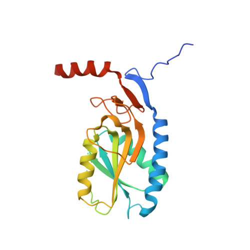
    6APS, 6APT, 6APU, 6APV, 6AQO, 6AR9

  • PubMed Abstract: 

    Due to toxicity and compliance issues and the emergence of resistance to current medications new drugs for the treatment of Human African Trypanosomiasis are needed. A potential approach to developing novel anti-trypanosomal drugs is by inhibition of the 6-oxopurine salvage pathways which synthesise the nucleoside monophosphates required for DNA/RNA production. This is in view of the fact that trypanosomes lack the machinery for de novo synthesis of the purine ring. To provide validation for this approach as a drug target, we have RNAi silenced the three 6-oxopurine phosphoribosyltransferase (PRTase) isoforms in the infectious stage of Trypanosoma brucei demonstrating that the combined activity of these enzymes is critical for the parasites' viability. Furthermore, we have determined crystal structures of two of these isoforms in complex with several acyclic nucleoside phosphonates (ANPs), a class of compound previously shown to inhibit 6-oxopurine PRTases from several species including Plasmodium falciparum. The most potent of these compounds have Ki values as low as 60 nM, and IC50 values in cell based assays as low as 4 μM. This data provides a solid platform for further investigations into the use of this pathway as a target for anti-trypanosomal drug discovery.


  • Organizational Affiliation
    • Institute of Parasitology, Biology Centre, Czech Academy of Sciences, Branišovská, České Budějovice, Czech Republic.

Macromolecule Content 

  • Total Structure Weight: 49.9 kDa 
  • Atom Count: 3,435 
  • Modeled Residue Count: 364 
  • Deposited Residue Count: 432 
  • Unique protein chains: 1

Macromolecules

Find similar proteins by:|  3D Structure
Entity ID: 1
MoleculeChains  Sequence LengthOrganismDetailsImage
Hypoxanthine-guanine phosphoribosyltransferase
A, B
216Trypanosoma brucei bruceiMutation(s): 0 
Gene Names: HGPRT
EC: 2.4.2.8
UniProt
Find proteins for Q07010 (Trypanosoma brucei brucei)
Explore Q07010 
Go to UniProtKB:  Q07010
Entity Groups
Sequence Clusters30% Identity50% Identity70% Identity90% Identity95% Identity100% Identity
UniProt GroupQ07010
Sequence Annotations
Expand
Reference Sequence

Small Molecules

Ligands 5 Unique
IDChains Name / Formula / InChI Key2D Diagram3D Interactions
SV2

Query on SV2


Download:Ideal Coordinates CCD File
C [auth A],
H [auth B]
[{2-[(guanine-9-yl)methyl]propane-1,3-diyl}bis(oxymethylene)]bis(phosphonic acid)
C11 H19 N5 O9 P2
VGXYLSOVNUSSKA-UHFFFAOYSA-N
TRS

Query on TRS


Download:Ideal Coordinates CCD File
F [auth A],
K [auth B]
2-AMINO-2-HYDROXYMETHYL-PROPANE-1,3-DIOL
C4 H12 N O3
LENZDBCJOHFCAS-UHFFFAOYSA-O
PEG

Query on PEG


Download:Ideal Coordinates CCD File
E [auth A],
J [auth B]
DI(HYDROXYETHYL)ETHER
C4 H10 O3
MTHSVFCYNBDYFN-UHFFFAOYSA-N
SO4

Query on SO4


Download:Ideal Coordinates CCD File
G [auth A]SULFATE ION
O4 S
QAOWNCQODCNURD-UHFFFAOYSA-L
MG

Query on MG


Download:Ideal Coordinates CCD File
D [auth A],
I [auth B]
MAGNESIUM ION
Mg
JLVVSXFLKOJNIY-UHFFFAOYSA-N

Experimental Data & Validation

Experimental Data

  • Method: X-RAY DIFFRACTION
  • Resolution: 1.76 Å
  • R-Value Free:  0.192 (Depositor), 0.193 (DCC) 
  • R-Value Work:  0.158 (Depositor), 0.158 (DCC) 
  • R-Value Observed: 0.160 (Depositor) 
Space Group: C 1 2 1
Unit Cell:
Length ( Å )Angle ( ˚ )
a = 116.013α = 90
b = 93.885β = 107.8
c = 44.715γ = 90
Software Package:
Software NamePurpose
PHENIXrefinement
XDSdata reduction
XDSdata scaling
PHASERphasing

Structure Validation

View Full Validation Report


Ligand Structure Quality Assessment 


Entry History 

Deposition Data

Revision History  (Full details and data files)

  • Version 1.0: 2018-03-14
    Type: Initial release
  • Version 1.1: 2023-10-04
    Changes: Data collection, Database references, Derived calculations, Refinement description