6AYR

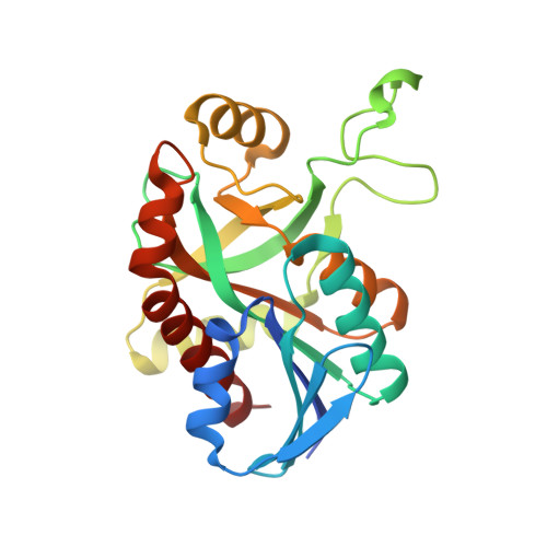
Crystal structure of Campylobacter jejuni 5'-methylthioadenosine/S-adenosyl homocysteine nucleosidase (MTAN) complexed with butylthio-DADMe-Immucillin-A


Experimental Data Snapshot

  • Method: X-RAY DIFFRACTION
  • Resolution: 1.95 Å
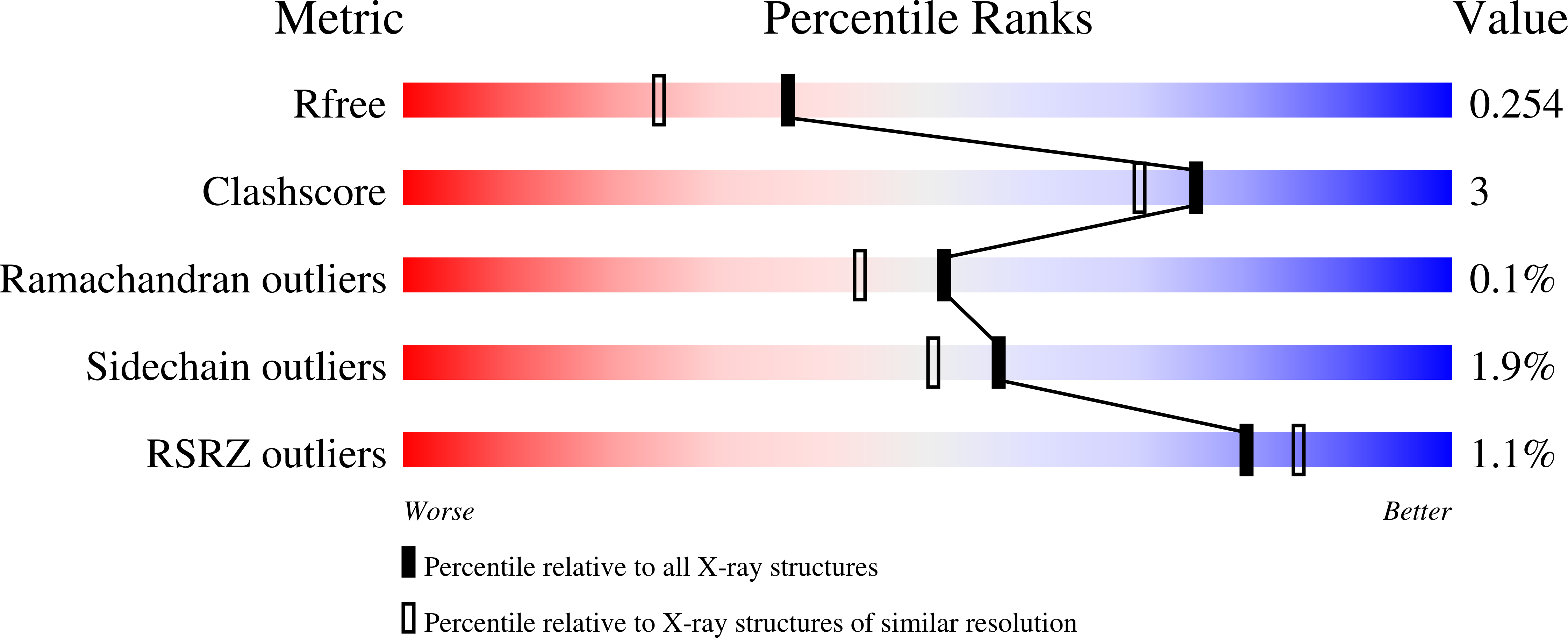
  • R-Value Free: 0.253 
  • R-Value Work: 0.223 
  • R-Value Observed: 0.225 

Starting Model: experimental
View more details

wwPDB Validation   3D Report Full Report


Ligand Structure Quality Assessment 


This is version 1.3 of the entry. See complete history


Literature

Transition-State Analogues of Campylobacter jejuni 5'-Methylthioadenosine Nucleosidase.

Ducati, R.G.Harijan, R.K.Cameron, S.A.Tyler, P.C.Evans, G.B.Schramm, V.L.

(2018) ACS Chem Biol 13: 3173-3183

  • DOI: https://doi.org/10.1021/acschembio.8b00781
  • Primary Citation of Related Structures:  
    6AYM, 6AYO, 6AYQ, 6AYR, 6AYS, 6AYT

  • PubMed Abstract: 

    Campylobacter jejuni is a Gram-negative bacterium responsible for food-borne gastroenteritis and associated with Guillain-Barré, Reiter, and irritable bowel syndromes. Antibiotic resistance in C. jejuni is common, creating a need for antibiotics with novel mechanisms of action. Menaquinone biosynthesis in C. jejuni uses the rare futalosine pathway, where 5'-methylthioadenosine nucleosidase ( CjMTAN) is proposed to catalyze the essential hydrolysis of adenine from 6-amino-6-deoxyfutalosine to form dehypoxanthinylfutalosine, a menaquinone precursor. The substrate specificity of CjMTAN is demonstrated to include 6-amino-6-deoxyfutalosine, 5'-methylthioadenosine, S-adenosylhomocysteine, adenosine, and 5'-deoxyadenosine. These activities span the catalytic specificities for the role of bacterial MTANs in menaquinone synthesis, quorum sensing, and S-adenosylmethionine recycling. We determined inhibition constants for potential transition-state analogues of CjMTAN. The best of these compounds have picomolar dissociation constants and were slow-onset tight-binding inhibitors. The most potent CjMTAN transition-state analogue inhibitors inhibited C. jejuni growth in culture at low micromolar concentrations, similar to gentamicin. The crystal structure of apoenzyme C. jejuni MTAN was solved at 1.25 Å, and five CjMTAN complexes with transition-state analogues were solved at 1.42 to 1.95 Å resolution. Inhibitor binding induces a loop movement to create a closed catalytic site with Asp196 and Ile152 providing purine leaving group activation and Arg192 and Glu12 activating the water nucleophile. With inhibitors bound, the interactions of the 4'-alkylthio or 4'-alkyl groups of this inhibitor family differ from the Escherichia coli MTAN structure by altered protein interactions near the hydrophobic pocket that stabilizes 4'-substituents of transition-state analogues. These CjMTAN inhibitors have potential as specific antibiotic candidates against C. jejuni.


  • Organizational Affiliation

    Department of Biochemistry , Albert Einstein College of Medicine , Bronx , New York 10461 , United States.


Macromolecules
Find similar proteins by:  (by identity cutoff)  |  3D Structure
Entity ID: 1
MoleculeChains Sequence LengthOrganismDetailsImage
5'-methylthioadenosine/S-adenosylhomocysteine nucleosidase
A, B, C, D
238Campylobacter jejuniMutation(s): 0 
Gene Names: A0M70_07260AJY53_05400
EC: 3.2.2.9 (PDB Primary Data), 3.2.2.30 (UniProt)
UniProt
Find proteins for Q0PC20 (Campylobacter jejuni subsp. jejuni serotype O:2 (strain ATCC 700819 / NCTC 11168))
Explore Q0PC20 
Go to UniProtKB:  Q0PC20
Entity Groups  
Sequence Clusters30% Identity50% Identity70% Identity90% Identity95% Identity100% Identity
UniProt GroupQ0PC20
Sequence Annotations
Expand
  • Reference Sequence
Small Molecules
Ligands 2 Unique
IDChains Name / Formula / InChI Key2D Diagram3D Interactions
BIG
Query on BIG

Download Ideal Coordinates CCD File 
E [auth A],
G [auth B],
H [auth C],
J [auth D]
(3R,4S)-1-[(4-amino-5H-pyrrolo[3,2-d]pyrimidin-7-yl)methyl]-4-[(butylsulfanyl)methyl]pyrrolidin-3-ol
C16 H25 N5 O S
LTSUEVPGSXUJHT-OLZOCXBDSA-N
EDO
Query on EDO

Download Ideal Coordinates CCD File 
F [auth A],
I [auth C],
K [auth D]
1,2-ETHANEDIOL
C2 H6 O2
LYCAIKOWRPUZTN-UHFFFAOYSA-N
Experimental Data & Validation

Experimental Data

  • Method: X-RAY DIFFRACTION
  • Resolution: 1.95 Å
  • R-Value Free: 0.253 
  • R-Value Work: 0.223 
  • R-Value Observed: 0.225 
  • Space Group: P 1 21 1
Unit Cell:
Length ( Å )Angle ( ˚ )
a = 68.156α = 90
b = 91.407β = 109.97
c = 78.431γ = 90
Software Package:
Software NamePurpose
REFMACrefinement
XDSdata reduction
Aimlessdata scaling
PHASERphasing

Structure Validation

View Full Validation Report


Ligand Structure Quality Assessment 


Entry History 

Deposition Data

Revision History  (Full details and data files)

  • Version 1.0: 2018-09-12
    Type: Initial release
  • Version 1.1: 2018-10-31
    Changes: Data collection, Database references
  • Version 1.2: 2018-11-28
    Changes: Data collection, Database references
  • Version 1.3: 2023-10-04
    Changes: Data collection, Database references, Refinement description