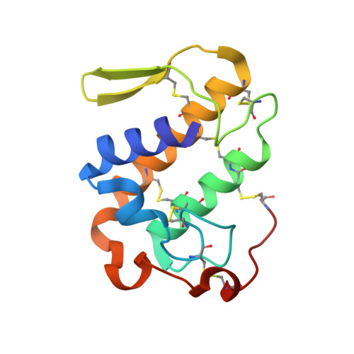
6B80

Crystal structure of myotoxin II from Bothrops moojeni complexed to myristic acid


Experimental Data Snapshot

  • Method: X-RAY DIFFRACTION
  • Resolution: 1.95 Å
  • R-Value Free: 0.198 
  • R-Value Work: 0.170 
  • R-Value Observed: 0.173 

Starting Model: experimental
View more details

wwPDB Validation   3D Report Full Report


Ligand Structure Quality Assessment 


This is version 1.4 of the entry. See complete history


Literature

Structural evidence for a fatty acid-independent myotoxic mechanism for a phospholipase A2-like toxin.

Salvador, G.H.M.Dos Santos, J.I.Borges, R.J.Fontes, M.R.M.

(2018) Biochim Biophys Acta Proteins Proteom 1866: 473-481

  • DOI: https://doi.org/10.1016/j.bbapap.2017.12.008
  • Primary Citation of Related Structures:  
    6B80, 6B81, 6B83, 6B84

  • PubMed Abstract: 

    The myotoxic mechanism for PLA 2 -like toxins has been proposed recently to be initiated by an allosteric change induced by a fatty acid binding to the protein, leading to the alignment of the membrane docking site (MDoS) and membrane disrupting site (MDiS). Previous structural studies performed by us demonstrated that MjTX-II, a PLA 2 -like toxin isolated from Bothrops moojeni, presents a different mode of ligand-interaction caused by natural amino acid substitutions and an insertion. Herein, we present four crystal structures of MjTX-II, in its apo state and complexed with fatty acids of different lengths. Analyses of these structures revealed slightly different oligomeric conformations but with both MDoSs in an arrangement that resembles an active-state PLA 2 -like structure. To explore the structural transitions between apo protein and fatty-acid complexes, we performed Normal Mode Molecular Dynamics simulations, revealing that oligomeric conformations of MjTX-II/fatty acid complexes may be reached in solution by the apo structure. Similar simulations with typical PLA 2 -like structures demonstrated that this transition is not possible without the presence of fatty acids. Thus, we hypothesize that MjTX-II does not require fatty acids to be active, although these ligands may eventually help in its stabilization by the formation of hydrogen bonds. Therefore, these results complement previous findings for MjTX-II and help us understand its particular ligand-binding properties and, more importantly, its particular mechanism of action, with a possible impact on the design of structure-based inhibitors for PLA 2 -like toxins in general.


  • Organizational Affiliation

    Departamento de Física e Biofísica, Instituto de Biociências, Universidade Estadual Paulista (UNESP), Botucatu, SP, Brazil.


Macromolecules
Find similar proteins by:  (by identity cutoff)  |  3D Structure
Entity ID: 1
MoleculeChains Sequence LengthOrganismDetailsImage
Basic phospholipase A2 homolog 2
A, B
122Bothrops moojeniMutation(s): 0 
UniProt
Find proteins for Q9I834 (Bothrops moojeni)
Explore Q9I834 
Go to UniProtKB:  Q9I834
Entity Groups  
Sequence Clusters30% Identity50% Identity70% Identity90% Identity95% Identity100% Identity
UniProt GroupQ9I834
Sequence Annotations
Expand
  • Reference Sequence
Small Molecules
Ligands 3 Unique
IDChains Name / Formula / InChI Key2D Diagram3D Interactions
PE4
Query on PE4

Download Ideal Coordinates CCD File 
H [auth B]2-{2-[2-(2-{2-[2-(2-ETHOXY-ETHOXY)-ETHOXY]-ETHOXY}-ETHOXY)-ETHOXY]-ETHOXY}-ETHANOL
C16 H34 O8
PJWQOENWHPEPKI-UHFFFAOYSA-N
MYR
Query on MYR

Download Ideal Coordinates CCD File 
C [auth A],
G [auth B]
MYRISTIC ACID
C14 H28 O2
TUNFSRHWOTWDNC-UHFFFAOYSA-N
SO4
Query on SO4

Download Ideal Coordinates CCD File 
D [auth A]
E [auth A]
F [auth A]
I [auth B]
J [auth B]
D [auth A],
E [auth A],
F [auth A],
I [auth B],
J [auth B],
K [auth B]
SULFATE ION
O4 S
QAOWNCQODCNURD-UHFFFAOYSA-L
Experimental Data & Validation

Experimental Data

  • Method: X-RAY DIFFRACTION
  • Resolution: 1.95 Å
  • R-Value Free: 0.198 
  • R-Value Work: 0.170 
  • R-Value Observed: 0.173 
  • Space Group: P 21 21 21
Unit Cell:
Length ( Å )Angle ( ˚ )
a = 50.545α = 90
b = 62.795β = 90
c = 86.848γ = 90
Software Package:
Software NamePurpose
SCALEPACKdata scaling
PHENIXrefinement
PDB_EXTRACTdata extraction
HKL-2000data reduction
PHASERphasing
Cootmodel building

Structure Validation

View Full Validation Report


Ligand Structure Quality Assessment 


Entry History & Funding Information

Deposition Data


Funding OrganizationLocationGrant Number
Sao Paulo Research Foundation (FAPESP)Brazil2015/24167-7

Revision History  (Full details and data files)

  • Version 1.0: 2018-10-03
    Type: Initial release
  • Version 1.1: 2019-04-17
    Changes: Author supporting evidence, Data collection
  • Version 1.2: 2020-01-01
    Changes: Author supporting evidence
  • Version 1.3: 2023-10-04
    Changes: Data collection, Database references, Refinement description
  • Version 1.4: 2024-10-23
    Changes: Structure summary