6C6Q | pdb_00006c6q

Crystal Structure of the Murine Norovirus VP1 P Domain in complex with the CD300lf Receptor


Experimental Data Snapshot

  • Method: X-RAY DIFFRACTION
  • Resolution: 2.00 Å
  • R-Value Free: 
    0.222 (Depositor), 0.224 (DCC) 
  • R-Value Work: 
    0.182 (Depositor), 0.185 (DCC) 
  • R-Value Observed: 
    0.183 (Depositor) 

Starting Models: experimental
View more details

wwPDB Validation 3D Report Full Report

Validation slider image for 6C6Q

This is version 1.5 of the entry. See complete history

Literature

Structural basis for murine norovirus engagement of bile acids and the CD300lf receptor.

Nelson, C.A.Wilen, C.B.Dai, Y.N.Orchard, R.C.Kim, A.S.Stegeman, R.A.Hsieh, L.L.Smith, T.J.Virgin, H.W.Fremont, D.H.

(2018) Proc Natl Acad Sci U S A 115: E9201-E9210

  • DOI: https://doi.org/10.1073/pnas.1805797115
  • Primary Citation Related Structures: 
    6C6Q, 6C74, 6CRJ, 6E47, 6E48

  • PubMed Abstract: 

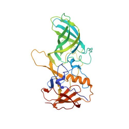
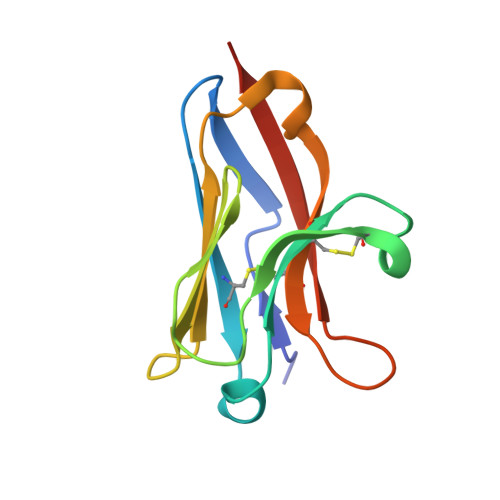
    Murine norovirus (MNoV) is closely related to human norovirus (HNoV), an infectious agent responsible for acute gastroenteritis worldwide. Here we report the X-ray crystal structure of the dimeric MNoV VP1 protruding (P) domain in complex with its cellular receptor CD300lf. CD300lf binds the P domain with a 2:2 stoichiometry, engaging a cleft between the AB and DE loops of the P2 subdomain at a site that overlaps the epitopes of neutralizing antibodies. We also identify that bile acids are cofactors enhancing MNoV cell-binding and infectivity. Structures of CD300lf-P domain in complex with glycochenodeoxycholic acid (GCDCA) and lithocholic acid (LCA) reveal two bile acid binding sites at the P domain dimer interface distant from receptor binding sites. The structural determinants for receptor and bile acid binding are supported by numerous biophysical assays utilizing interface residue mutations. We find that the monomeric affinity of CD300lf for the P domain is low and is divalent cation dependent. We have also determined the crystal structure of CD300lf in complex with phosphocholine, revealing that MNoV engages its receptor in a manner mimicking host ligands including similar metal coordination. Docking of the cocomplex structures onto a cryo-EM-derived model of MNoV suggests that each virion can make multiple CD300lf engagements, and thus, infection may be driven by the avidity of cell surface clustered CD300lf. These studies identify multiple potential modulators of norovirus infection that may act to regulate the interaction between the viral capsid P domain and its cognate cellular receptor.


  • Organizational Affiliation
    • Department of Pathology and Immunology, Washington University School of Medicine, St. Louis, MO 63110.

Macromolecule Content 

  • Total Structure Weight: 93.08 kDa 
  • Atom Count: 7,267 
  • Modeled Residue Count: 828 
  • Deposited Residue Count: 832 
  • Unique protein chains: 2

Macromolecules

Find similar proteins by:|  3D Structure
Entity ID: 1
MoleculeChains  Sequence LengthOrganismDetailsImage
Capsid protein VP1
A, B
303Murine norovirus 1Mutation(s): 0 
UniProt
Find proteins for Q80J94 (Norovirus (isolate Mouse/NoV/United States/MNV1/2002/GV))
Explore Q80J94 
Go to UniProtKB:  Q80J94
Entity Groups
Sequence Clusters30% Identity50% Identity70% Identity90% Identity95% Identity100% Identity
UniProt GroupQ80J94
Sequence Annotations
Expand
Reference Sequence
Find similar proteins by:|  3D Structure
Entity ID: 2
MoleculeChains  Sequence LengthOrganismDetailsImage
CMRF35-like molecule 1C [auth D],
D [auth F]
113Mus musculusMutation(s): 0 
Gene Names: Cd300lfClm1Lmir3
UniProt
Find proteins for Q6SJQ7 (Mus musculus)
Explore Q6SJQ7 
Go to UniProtKB:  Q6SJQ7
Entity Groups
Sequence Clusters30% Identity50% Identity70% Identity90% Identity95% Identity100% Identity
UniProt GroupQ6SJQ7
Sequence Annotations
Expand
Reference Sequence

Small Molecules

Ligands 3 Unique
IDChains Name / Formula / InChI Key2D Diagram3D Interactions
EDO

Query on EDO


Download:Ideal Coordinates CCD File
AA [auth D]
BA [auth D]
CA [auth D]
E [auth A]
EA [auth F]
AA [auth D],
BA [auth D],
CA [auth D],
E [auth A],
EA [auth F],
F [auth A],
FA [auth F],
G [auth A],
GA [auth F],
H [auth A],
I [auth A],
J [auth A],
K [auth A],
P [auth B],
Q [auth B],
R [auth B],
S [auth B],
T [auth B],
U [auth B],
X [auth D],
Y [auth D],
Z [auth D]
1,2-ETHANEDIOL
C2 H6 O2
LYCAIKOWRPUZTN-UHFFFAOYSA-N
CL

Query on CL


Download:Ideal Coordinates CCD File
L [auth A],
M [auth A]
CHLORIDE ION
Cl
VEXZGXHMUGYJMC-UHFFFAOYSA-M
MG

Query on MG


Download:Ideal Coordinates CCD File
DA [auth D]
HA [auth F]
N [auth A]
O [auth A]
V [auth B]
DA [auth D],
HA [auth F],
N [auth A],
O [auth A],
V [auth B],
W [auth B]
MAGNESIUM ION
Mg
JLVVSXFLKOJNIY-UHFFFAOYSA-N

Experimental Data & Validation

Experimental Data

  • Method: X-RAY DIFFRACTION
  • Resolution: 2.00 Å
  • R-Value Free:  0.222 (Depositor), 0.224 (DCC) 
  • R-Value Work:  0.182 (Depositor), 0.185 (DCC) 
  • R-Value Observed: 0.183 (Depositor) 
Space Group: P 21 21 21
Unit Cell:
Length ( Å )Angle ( ˚ )
a = 48.712α = 90
b = 135.245β = 90
c = 139.516γ = 90
Software Package:
Software NamePurpose
Aimlessdata scaling
PHASERphasing
PHENIXrefinement
PDB_EXTRACTdata extraction
XDSdata reduction

Structure Validation

View Full Validation Report


Entry History 

& Funding Information

Deposition Data


Funding OrganizationLocationGrant Number
National Institutes of Health/National Institute Of Allergy and Infectious Diseases (NIH/NIAID)United StatesAI109725
National Institutes of Health/National Institute Of Allergy and Infectious Diseases (NIH/NIAID)United StatesAI127552

Revision History  (Full details and data files)

  • Version 1.0: 2018-09-12
    Type: Initial release
  • Version 1.1: 2018-09-19
    Changes: Data collection, Database references
  • Version 1.2: 2018-10-03
    Changes: Data collection, Database references
  • Version 1.3: 2019-12-18
    Changes: Author supporting evidence
  • Version 1.4: 2023-10-04
    Changes: Data collection, Database references, Derived calculations, Refinement description
  • Version 1.5: 2024-10-30
    Changes: Structure summary