6DK1 | pdb_00006dk1

Human sigma-1 receptor bound to (+)-pentazocine


Experimental Data Snapshot

  • Method: X-RAY DIFFRACTION
  • Resolution: 3.12 Å
  • R-Value Free: 
    0.278 (Depositor), 0.277 (DCC) 
  • R-Value Work: 
    0.247 (Depositor), 0.249 (DCC) 
  • R-Value Observed: 
    0.248 (Depositor) 

Starting Model: experimental
View more details

wwPDB Validation 3D Report Full Report

Validation slider image for 6DK1

Ligand Structure Quality Assessment 


This is version 1.2 of the entry. See complete history

Literature

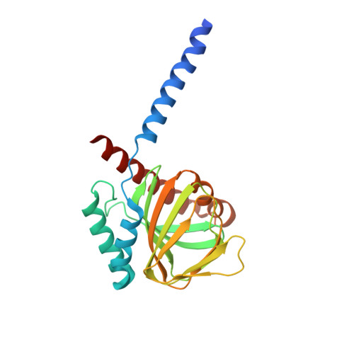
Structural basis for sigma1receptor ligand recognition.

Schmidt, H.R.Betz, R.M.Dror, R.O.Kruse, A.C.

(2018) Nat Struct Mol Biol 25: 981-987

  • DOI: https://doi.org/10.1038/s41594-018-0137-2
  • Primary Citation Related Structures: 
    6DJZ, 6DK0, 6DK1

  • PubMed Abstract: 

    The σ 1 receptor is a poorly understood membrane protein expressed throughout the human body. Ligands targeting the σ 1 receptor are in clinical trials for treatment of Alzheimer's disease, ischemic stroke, and neuropathic pain. However, relatively little is known regarding the σ 1 receptor's molecular function. Here, we present crystal structures of human σ 1 receptor bound to the antagonists haloperidol and NE-100, and the agonist (+)-pentazocine, at crystallographic resolutions of 3.1 Å, 2.9 Å, and 3.1 Å, respectively. These structures reveal a unique binding pose for the agonist. The structures and accompanying molecular dynamics (MD) simulations identify agonist-induced structural rearrangements in the receptor. Additionally, we show that ligand binding to σ 1 is a multistep process that is rate limited by receptor conformational change. We used MD simulations to reconstruct a ligand binding pathway involving two major conformational changes. These data provide a framework for understanding the molecular basis for σ 1 agonism.


  • Organizational Affiliation
    • Department of Biological Chemistry and Molecular Pharmacology, Harvard Medical School, Boston, MA, USA.

Macromolecule Content 

  • Total Structure Weight: 82.84 kDa 
  • Atom Count: 5,407 
  • Modeled Residue Count: 652 
  • Deposited Residue Count: 681 
  • Unique protein chains: 1

Macromolecules

Find similar proteins by:|  3D Structure
Entity ID: 1
MoleculeChains  Sequence LengthOrganismDetailsImage
Sigma non-opioid intracellular receptor 1
A, B, C
227Homo sapiensMutation(s): 0 
Gene Names: SIGMAR1OPRS1SRBPAAG8
Membrane Entity: Yes 
UniProt & NIH Common Fund Data Resources
Find proteins for Q99720 (Homo sapiens)
Explore Q99720 
Go to UniProtKB:  Q99720
PHAROS:  Q99720
GTEx:  ENSG00000147955 
Entity Groups
Sequence Clusters30% Identity50% Identity70% Identity90% Identity95% Identity100% Identity
UniProt GroupQ99720
Sequence Annotations
Expand
Reference Sequence

Small Molecules

Ligands 4 Unique
IDChains Name / Formula / InChI Key2D Diagram3D Interactions
OLC

Query on OLC


Download:Ideal Coordinates CCD File
DA [auth C]
EA [auth C]
FA [auth C]
GA [auth C]
HA [auth C]
DA [auth C],
EA [auth C],
FA [auth C],
GA [auth C],
HA [auth C],
IA [auth C],
M [auth A],
N [auth A],
U [auth B],
V [auth B],
W [auth B]
(2R)-2,3-dihydroxypropyl (9Z)-octadec-9-enoate
C21 H40 O4
RZRNAYUHWVFMIP-GDCKJWNLSA-N
GM4

Query on GM4


Download:Ideal Coordinates CCD File
D [auth A],
O [auth B],
X [auth C]
(2S,6S,11S)-6,11-dimethyl-3-(3-methylbut-2-en-1-yl)-1,2,3,4,5,6-hexahydro-2,6-methano-3-benzazocin-8-ol
C19 H27 N O
VOKSWYLNZZRQPF-CCKFTAQKSA-N
SO4

Query on SO4


Download:Ideal Coordinates CCD File
AA [auth C]
E [auth A]
F [auth A]
G [auth A]
H [auth A]
AA [auth C],
E [auth A],
F [auth A],
G [auth A],
H [auth A],
I [auth A],
J [auth A],
K [auth A],
P [auth B],
Q [auth B],
R [auth B],
S [auth B],
Y [auth C],
Z [auth C]
SULFATE ION
O4 S
QAOWNCQODCNURD-UHFFFAOYSA-L
GOL

Query on GOL


Download:Ideal Coordinates CCD File
BA [auth C],
CA [auth C],
L [auth A],
T [auth B]
GLYCEROL
C3 H8 O3
PEDCQBHIVMGVHV-UHFFFAOYSA-N

Experimental Data & Validation

Experimental Data

  • Method: X-RAY DIFFRACTION
  • Resolution: 3.12 Å
  • R-Value Free:  0.278 (Depositor), 0.277 (DCC) 
  • R-Value Work:  0.247 (Depositor), 0.249 (DCC) 
  • R-Value Observed: 0.248 (Depositor) 
Space Group: P 21 21 2
Unit Cell:
Length ( Å )Angle ( ˚ )
a = 84.837α = 90
b = 128.59β = 90
c = 109.18γ = 90
Software Package:
Software NamePurpose
PHENIXrefinement
XDSdata reduction
Aimlessdata scaling
PHENIXphasing

Structure Validation

View Full Validation Report


Ligand Structure Quality Assessment 


Entry History 

& Funding Information

Deposition Data


Funding OrganizationLocationGrant Number
National Science Foundation (NSF, United States)United StatesDGE1745303
National Institutes of Health/National Institute of General Medical Sciences (NIH/NIGMS)United StatesR01 GM119185

Revision History  (Full details and data files)

  • Version 1.0: 2018-10-17
    Type: Initial release
  • Version 1.1: 2019-11-27
    Changes: Author supporting evidence
  • Version 1.2: 2023-10-11
    Changes: Data collection, Database references, Refinement description