6F0A | pdb_00006f0a

Crystal structure of human indoleamine 2,3-dioxygenase bound to a triazole inhibitor and alanine molecule.


Experimental Data Snapshot

  • Method: X-RAY DIFFRACTION
  • Resolution: 2.26 Å
  • R-Value Free: 
    0.225 (Depositor), 0.233 (DCC) 
  • R-Value Work: 
    0.191 (Depositor), 0.199 (DCC) 
  • R-Value Observed: 
    0.193 (Depositor) 

Starting Model: experimental
View more details

wwPDB Validation 3D Report Full Report

Validation slider image for 6F0A

Ligand Structure Quality Assessment 


This is version 1.5 of the entry. See complete history

Literature

New 4-Amino-1,2,3-Triazole Inhibitors of Indoleamine 2,3-Dioxygenase Form a Long-Lived Complex with the Enzyme and Display Exquisite Cellular Potency.

Alexandre, J.A.C.Swan, M.K.Latchem, M.J.Boyall, D.Pollard, J.R.Hughes, S.W.Westcott, J.

(2018) Chembiochem 19: 552-561

  • DOI: https://doi.org/10.1002/cbic.201700560
  • Primary Citation Related Structures: 
    6F0A

  • PubMed Abstract: 

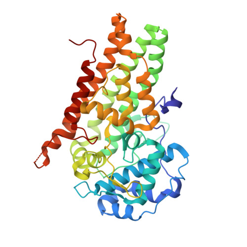
    Indoleamine-2,3 dioxygenase 1 (IDO1) has emerged as a central regulator of immune responses in both normal and disease biology. Due to its established role in promoting tumour immune escape, IDO1 has become an attractive target for cancer treatment. A novel series of highly cell potent IDO1 inhibitors based on a 4-amino-1,2,3-triazole core have been identified. Comprehensive kinetic, biochemical and structural studies demonstrate that compounds from this series have a noncompetitive kinetic mechanism of action with respect to the tryptophan substrate. In co-complex crystal structures, the compounds bind in the tryptophan pocket and make a direct ligand interaction with the haem iron of the porphyrin cofactor. It is proposed that these data can be rationalised by an ordered-binding mechanism, in which the inhibitor binds an apo form of the enzyme that is not competent to bind tryptophan. These inhibitors also form a very tight, long-lived complex with the enzyme, which partially explains their exquisite cellular potency. This novel series represents an attractive starting point for the future development of potent IDO1-targeted drugs.


  • Organizational Affiliation
    • Vertex Pharmaceuticals (Europe) Limited, 86-88 Jubilee Avenue, Abingdon, Oxfordshire, OX14 4RW, UK.

Macromolecule Content 

  • Total Structure Weight: 90.25 kDa 
  • Atom Count: 6,364 
  • Modeled Residue Count: 764 
  • Deposited Residue Count: 786 
  • Unique protein chains: 1

Macromolecules

Find similar proteins by:|  3D Structure
Entity ID: 1
MoleculeChains  Sequence LengthOrganismDetailsImage
Indoleamine 2,3-dioxygenase 1A,
B [auth C]
393Homo sapiensMutation(s): 0 
Gene Names: IDO1IDOINDO
EC: 1.13.11.52
UniProt & NIH Common Fund Data Resources
Find proteins for P14902 (Homo sapiens)
Explore P14902 
Go to UniProtKB:  P14902
PHAROS:  P14902
GTEx:  ENSG00000131203 
Entity Groups
Sequence Clusters30% Identity50% Identity70% Identity90% Identity95% Identity100% Identity
UniProt GroupP14902
Sequence Annotations
Expand
Reference Sequence

Small Molecules

Ligands 3 Unique
IDChains Name / Formula / InChI Key2D Diagram3D Interactions
HEM

Query on HEM


Download:Ideal Coordinates CCD File
E [auth A],
H [auth C]
PROTOPORPHYRIN IX CONTAINING FE
C34 H32 Fe N4 O4
KABFMIBPWCXCRK-RGGAHWMASA-L
C82
(Subject of Investigation/LOI)

Query on C82


Download:Ideal Coordinates CCD File
C [auth A],
F [auth C]
~{N}-(4-chlorophenyl)-1~{H}-1,2,3-triazol-5-amine
C8 H7 Cl N4
IUTZKZLVPUPHDA-UHFFFAOYSA-N
ALA

Query on ALA


Download:Ideal Coordinates CCD File
D [auth A],
G [auth C]
ALANINE
C3 H7 N O2
QNAYBMKLOCPYGJ-REOHCLBHSA-N

Experimental Data & Validation

Experimental Data

  • Method: X-RAY DIFFRACTION
  • Resolution: 2.26 Å
  • R-Value Free:  0.225 (Depositor), 0.233 (DCC) 
  • R-Value Work:  0.191 (Depositor), 0.199 (DCC) 
  • R-Value Observed: 0.193 (Depositor) 
Space Group: P 21 21 21
Unit Cell:
Length ( Å )Angle ( ˚ )
a = 85.48α = 90
b = 96.89β = 90
c = 132.19γ = 90
Software Package:
Software NamePurpose
xia2data scaling
BUSTERrefinement
PDB_EXTRACTdata extraction
xia2data reduction
MOLREPphasing

Structure Validation

View Full Validation Report


Ligand Structure Quality Assessment 


Entry History 

Deposition Data

Revision History  (Full details and data files)

  • Version 1.0: 2017-12-27
    Type: Initial release
  • Version 1.1: 2018-02-07
    Changes: Database references
  • Version 1.2: 2018-04-04
    Changes: Data collection, Database references
  • Version 1.3: 2019-10-16
    Changes: Data collection
  • Version 1.4: 2024-01-17
    Changes: Data collection, Database references, Derived calculations, Refinement description
  • Version 1.5: 2024-11-06
    Changes: Structure summary