6FSF | pdb_00006fsf

Crystal structure of the tandem PX-PH-domains of Bem3 from Saccharomyces cerevisiae


Experimental Data Snapshot

  • Method: X-RAY DIFFRACTION
  • Resolution: 2.20 Å
  • R-Value Free: 
    0.234 (Depositor), 0.241 (DCC) 
  • R-Value Work: 
    0.211 (Depositor), 0.217 (DCC) 
  • R-Value Observed: 
    0.212 (Depositor) 

wwPDB Validation 3D Report Full Report

Validation slider image for 6FSF

This is version 1.2 of the entry. See complete history

Literature

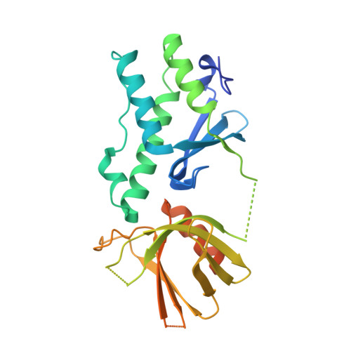
Structure of the tandem PX-PH domains of Bem3 from Saccharomyces cerevisiae.

Ali, I.Eu, S.Koch, D.Bleimling, N.Goody, R.S.Muller, M.P.

(2018) Acta Crystallogr F Struct Biol Commun 74: 315-321

  • DOI: https://doi.org/10.1107/S2053230X18005915
  • Primary Citation Related Structures: 
    6FSF

  • PubMed Abstract: 

    The structure of the tandem lipid-binding PX and pleckstrin-homology (PH) domains of the Cdc42 GTPase-activating protein Bem3 from Saccharomyces cerevisiae (strain S288c) has been determined to a resolution of 2.2 Å (R work = 21.1%, R free = 23.4%). It shows that the domains adopt a relative orientation that enables them to simultaneously bind to a membrane and suggests possible cooperativity in membrane binding.


  • Organizational Affiliation
    • Department of Structural Biochemistry, Max Planck Institute of Molecular Physiology, Otto-Hahn-Strasse 11, 44227 Dortmund, Germany.

Macromolecule Content 

  • Total Structure Weight: 30.74 kDa 
  • Atom Count: 1,808 
  • Modeled Residue Count: 218 
  • Deposited Residue Count: 269 
  • Unique protein chains: 1

Macromolecules

Find similar proteins by:|  3D Structure
Entity ID: 1
MoleculeChains  Sequence LengthOrganismDetailsImage
GTPase-activating protein BEM3269Saccharomyces cerevisiae S288CMutation(s): 0 
Gene Names: BEM3YPL115CLPH12C
UniProt
Find proteins for P32873 (Saccharomyces cerevisiae (strain ATCC 204508 / S288c))
Explore P32873 
Go to UniProtKB:  P32873
Entity Groups
Sequence Clusters30% Identity50% Identity70% Identity90% Identity95% Identity100% Identity
UniProt GroupP32873
Sequence Annotations
Expand
Reference Sequence

Experimental Data & Validation

Experimental Data

  • Method: X-RAY DIFFRACTION
  • Resolution: 2.20 Å
  • R-Value Free:  0.234 (Depositor), 0.241 (DCC) 
  • R-Value Work:  0.211 (Depositor), 0.217 (DCC) 
  • R-Value Observed: 0.212 (Depositor) 
Space Group: P 62
Unit Cell:
Length ( Å )Angle ( ˚ )
a = 85.37α = 90
b = 85.37β = 90
c = 63.97γ = 120
Software Package:
Software NamePurpose
PHENIXrefinement
XDSdata reduction
XSCALEdata scaling
PHENIXphasing

Structure Validation

View Full Validation Report


Entry History 

& Funding Information

Deposition Data


Funding OrganizationLocationGrant Number
German Research FoundationGermanyDFG/ANR grant GO 284/8-1
German Research FoundationGermanySFB 642

Revision History  (Full details and data files)

  • Version 1.0: 2018-05-02
    Type: Initial release
  • Version 1.1: 2018-05-09
    Changes: Data collection, Database references
  • Version 1.2: 2024-05-08
    Changes: Data collection, Database references